Review
. 2013 Mar 10;18(8):899-929.
doi: 10.1089/ars.2012.4795. Epub 2012 Oct 12.

Oxidative stress, redox signaling, and metal chelation in anthracycline cardiotoxicity and pharmacological cardioprotection

Affiliations
Review

Oxidative stress, redox signaling, and metal chelation in anthracycline cardiotoxicity and pharmacological cardioprotection

Martin Stěrba et al. Antioxid Redox Signal. .

Abstract

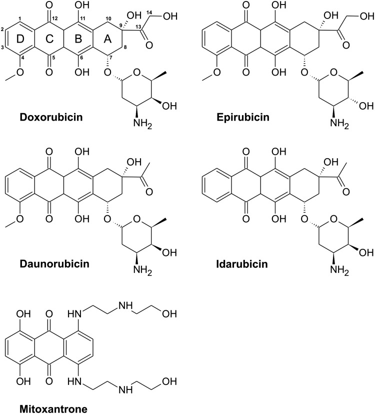
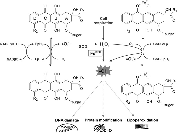
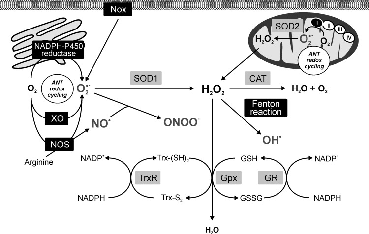
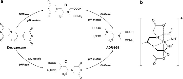
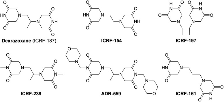
Significance: Anthracyclines (doxorubicin, daunorubicin, or epirubicin) rank among the most effective anticancer drugs, but their clinical usefulness is hampered by the risk of cardiotoxicity. The most feared are the chronic forms of cardiotoxicity, characterized by irreversible cardiac damage and congestive heart failure. Although the pathogenesis of anthracycline cardiotoxicity seems to be complex, the pivotal role has been traditionally attributed to the iron-mediated formation of reactive oxygen species (ROS). In clinics, the bisdioxopiperazine agent dexrazoxane (ICRF-187) reduces the risk of anthracycline cardiotoxicity without a significant effect on response to chemotherapy. The prevailing concept describes dexrazoxane as a prodrug undergoing bioactivation to an iron-chelating agent ADR-925, which may inhibit anthracycline-induced ROS formation and oxidative damage to cardiomyocytes.

Recent advances: A considerable body of evidence points to mitochondria as the key targets for anthracycline cardiotoxicity, and therefore it could be also crucial for effective cardioprotection. Numerous antioxidants and several iron chelators have been tested in vitro and in vivo with variable outcomes. None of these compounds have matched or even surpassed the effectiveness of dexrazoxane in chronic anthracycline cardiotoxicity settings, despite being stronger chelators and/or antioxidants.

Critical issues: The interpretation of many findings is complicated by the heterogeneity of experimental models and frequent employment of acute high-dose treatments with limited translatability to clinical practice.

Future directions: Dexrazoxane may be the key to the enigma of anthracycline cardiotoxicity, and therefore it warrants further investigation, including the search for alternative/complementary modes of cardioprotective action beyond simple iron chelation.


Figures

FIG. 1.
FIG. 1.
Chemical structures of the most important anthracycline antibiotics and mitoxantrone.
FIG. 2.
FIG. 2.
Overview of molecular changes associated with development of anthracycline cardiotoxicity. Green color indicates increase, whereas red color illustrates decrease due to the treatment. A, anthracycline; CK, creatine kinase; Cr, creatine; Cyt c, cytochrome c; MLC, myosin light chains 1 and 2; MMPs, metalloproteinases; PCr, phosphocreatine; RyR, ryanodine receptor; SERCA, sarco-/endoplasmic reticulum Ca2+-ATPase; SR, sarco-/endoplasmic reticulum; ROS, reactive oxygen species; TOP2, topoisomerase II; VIM, vimentin; UBI, ubiquitin. (To see this illustration in color, the reader is referred to the web version of this article at www.liebertpub.com/ars.)
FIG. 3.
FIG. 3.
Schematic illustration of main pathways of anthracycline-induced and iron-catalyzed oxidative stress production. Fe, iron; Fp, flavoprotein; GSH, reduced glutathione; GSSG, oxidized glutathione; H2O2, hydrogen peroxide; NAD(P), nicotinamide adenine dinucleotide (phosphate); •O2, superoxide radical; •OH, hydroxyl radical; SOD, superoxide dismutase.
FIG. 4.
FIG. 4.
Anthracycline-induced reactive oxygen/nitrogen species production within the cell context. Black panels—anthracycline redox-cycling sites; gray panels—antioxidant defense. I, II, III, and IV, respiratory chain complexes; ANT, anthracyclines; CAT, catalase; GPx, glutathione peroxidase; GR, glutathione reductase; GSH, reduced glutathione; GSSG, oxidized glutathione; H2O2, hydrogen peroxide; NAD(P), nicotinamide adenine dinucleotide (phosphate); NO•, NO radical; NOS, NO synthase; Nox, NADPH oxidase; •O2, superoxide radical; •OH, hydroxyl radical; ONOO, peroxynitrite; SOD1, cytosolic superoxide dismutase (CuZnSOD); SOD2, mitochondrial superoxide dismutase (MnSOD); TrxR, thioredoxinreductase; Trx-S2, oxidized thioredoxin; Trx-(SH)2, reduced thioredoxin; XO, xanthine oxidase.
FIG. 5.
FIG. 5.
Schematic overview of mitochondrial alterations associated with the anthracycline cardiotoxicity. Green color indicates increase, whereas red color illustrates decrease due to the treatment. I, II, III, and IV, respiratory chain complexes; A, anthracycline; A•, anthracycline semiquinone radical; β-ox., β-oxidation; ANT, adenine nucleotide translocase-1; c, cardiolipin; c, cytochrome c; smtCK, sarcomeric mitochondrial creatine kinase; Cr, creatine; F0 and F1, ATP-synthase (complex V) domains; FA, fatty acids; FABP, fatty acid-binding proteins; MCU, mitochondrial calcium uniporter; MnSOD, mitochondrial isoform of superoxide dismutase; mtDNA, mitochondrial DNA; mPTP, mitochondrial permeability transition pore; NAD, nicotinamide adenine dinucleotide; P, phosphorylation; PCr, phosphocreatine; PDE1, pyruvate dehydrogenase-1; Q, coenzyme Q; RISP, Reiske iron–sulfur protein; ROS, reactive oxygen species; TCA cycle, tricarboxylic acid cycle; TOP2, topoisomerase 2; VDAC, voltage-dependent anion channel-1. (To see this illustration in color, the reader is referred to the web version of this article at www.liebertpub.com/ars.)
FIG. 6.
FIG. 6.
Chemistry of dexrazoxane (ICRF-187). (a) Stepwise hydrolysis of dexrazoxane to intermediate metabolites B and C and iron-chelating metabolite ADR-925. (b) Chemical structure of complexes of ADR-925 with Fe3+.
FIG. 7.
FIG. 7.
Chemical structures of selected bisdioxopiperazine derivatives tested for their cardioprotective effects against anthracycline cardiotoxicity.
FIG. 8.
FIG. 8.
Chemical structures of iron chelators tested for their cardioprotective effects against anthracycline cardiotoxicity.
FIG. 9.
FIG. 9.
Schematic illustration of anthracycline-induced changes in iron metabolism. DMT1, divalent metal transporter 1; FPN, ferroportin 1; Mfrn, mitoferrin; MtF, mitochondrial ferritin; STEAP, six-transmembrane epithelial antigen of prostate 3; Tf, transferrin; TfR, transferrin receptor. (To see this illustration in color, the reader is referred to the web version of this article at www.liebertpub.com/ars.)

References

    1. Adachi K. Fujiura Y. Mayumi F. Nozuhara A. Sugiu Y. Sakanashi T. Hidaka T. Toshima H. A deletion of mitochondrial DNA in murine doxorubicin-induced cardiotoxicity. Biochem Biophys Res Commun. 1993;195:945–951. - PubMed
    1. Adams MJ. Lipshultz SE. Pathophysiology of anthracycline- and radiation-associated cardiomyopathies: implications for screening and prevention. Pediatr Blood Cancer. 2005;44:600–606. - PubMed
    1. al-Harbi MM. al-Gharably NM. al-Shabanah OA. al-Bekairi AM. Osman AM. Tawfik HN. Prevention of doxorubicin-induced myocardial and haematological toxicities in rats by the iron chelator desferrioxamine. Cancer Chemother Pharmacol. 1992;31:200–204. - PubMed
    1. Al-Rousan RM. Paturi S. Laurino JP. Kakarla SK. Gutta AK. Walker EM. Blough ER. Deferasirox removes cardiac iron and attenuates oxidative stress in the iron-overloaded gerbil. Am J Hematol. 2009;84:565–570. - PubMed
    1. Alderton P. Gross J. Green MD. Role of (+-)-1,2-bis(3,5-dioxopiperazinyl-1-yl)propane (ICRF-187) in modulating free radical scavenging enzymes in doxorubicin-induced cardiomyopathy. Cancer Res. 1990;50:5136–5142. - PubMed

Publication types

MeSH terms