. 2012 Jul 12;75(1):94-107.
doi: 10.1016/j.neuron.2012.04.033.

Distinct roles of muscle and motoneuron LRP4 in neuromuscular junction formation

Affiliations

Distinct roles of muscle and motoneuron LRP4 in neuromuscular junction formation

Haitao Wu et al. Neuron. .

Erratum in

  • Neuron. 2012 Sep 6;75(5):930

Abstract

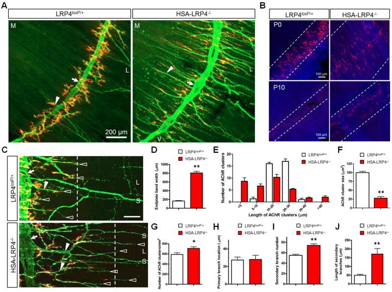
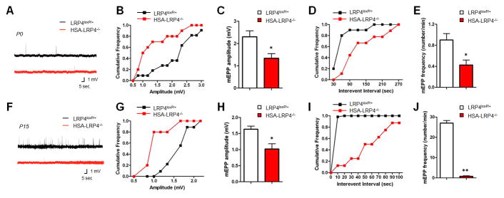
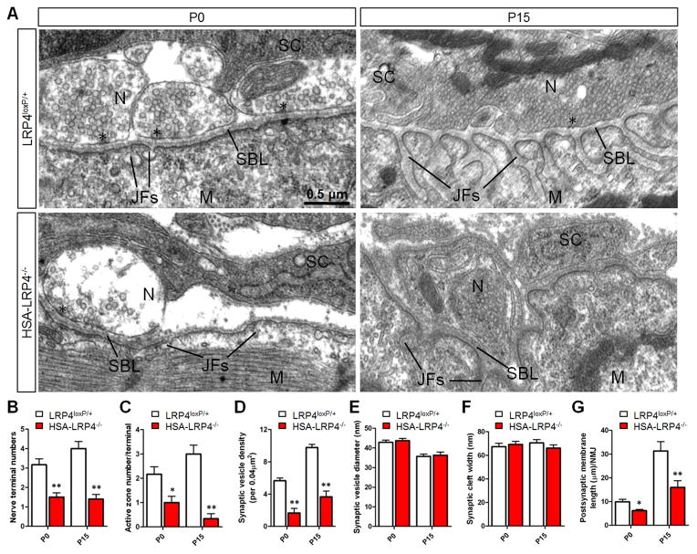
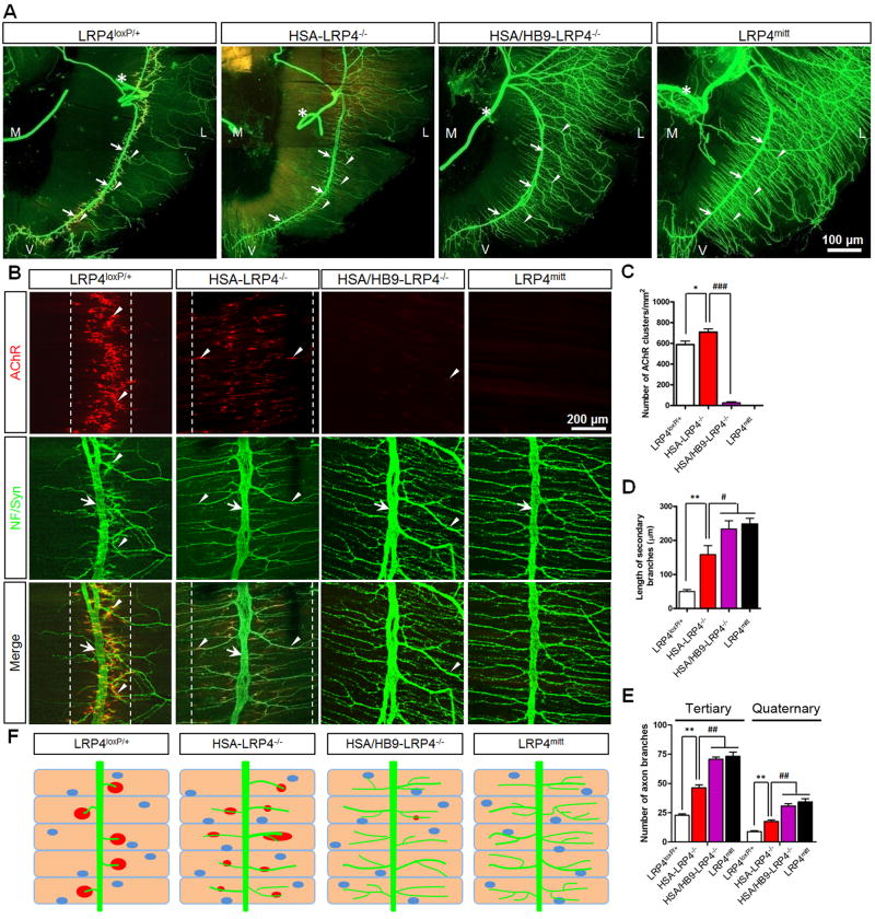
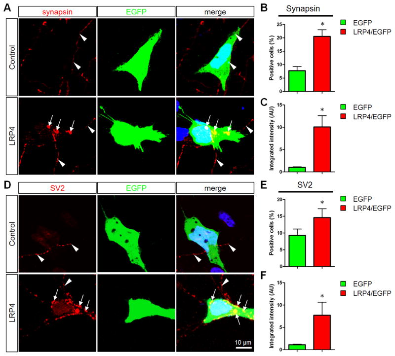
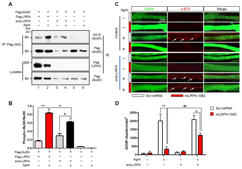
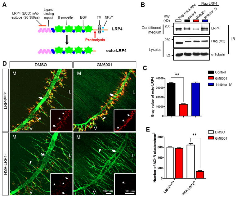
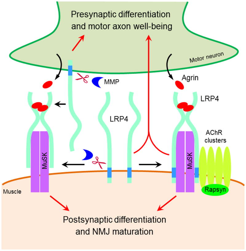
Neuromuscular junction (NMJ) formation requires precise interaction between motoneurons and muscle fibers. LRP4 is a receptor of agrin that is thought to act in cis to stimulate MuSK in muscle fibers for postsynaptic differentiation. Here we dissected the roles of LRP4 in muscle fibers and motoneurons in NMJ formation by cell-specific mutation. Studies of muscle-specific mutants suggest that LRP4 is involved in deciding where to form AChR clusters in muscle fibers, postsynaptic differentiation, and axon terminal development. LRP4 in HEK293 cells increased synapsin or SV2 puncta in contacting axons of cocultured neurons, suggesting a synaptogenic function. Analysis of LRP4 muscle and motoneuron double mutants and mechanistic studies suggest that NMJ formation may also be regulated by LRP4 in motoneurons, which could serve as agrin's receptor in trans to induce AChR clusters. These observations uncovered distinct roles of LRP4 in motoneurons and muscles in NMJ development.


Figures

Figure 1
Figure 1. Aberrant NMJ Formation in HSA-LRP4−/− Mice
(A) Abnormal NMJ morphology in HSA-LRP4−/− mice. P0 control and mutant diaphragms were stained whole-mount with α-BTX (red) to label AChR clusters and with NF/synaptophysin antibodies (Syn) to label nerve terminals, which was visualized by Alexa Fluor 488-conjugated goat anti-rabbit antibody (green). Shown were the left, ventral areas of hemi-diaphragms. Arrow, primary nerve branches; arrowheads, secondary nerve branches. M, medial; L, lateral; V, ventral. (B) Wider endplate areas in P0 and P10 HSA-LRP4−/− muscles, which were counterstained with phalloidin. Dashed lines indicate clusters-enriched regions. (C) Increased secondary nerve branches (arrowhead) and over-shot axon terminals (empty arrowheads). Arrows, primary branches; dashed lines, boundary of cluster-rich regions. S, sensory or autonomic nerve axons; L, lateral. (D–J) Compared to controls, HSA-LRP4−/− mice showed increased band width (**, P<0.01; n=5, t-test) (D), increased variability in cluster sizes (calculated using LSM Image Browser software, Zeiss) (E); decreased size of all clusters (F), increased number of AChR clusters (G); no difference in the distances between primary nerve branches and the muscle midlines (H); increased number of secondary/intramuscular branches (I); and increased length of secondary branches (J). Data are shown as mean ± SEM. *p < 0.05; **p < 0.01 (n = 4–8; t test). Please also see Figures S1 and S2.
Figure 2
Figure 2. Compromised NMJ Transmission in HSA-LRP4−/− Mice
mEPPs were recorded in P0 (A–E) and P15 (F–J) control and HSA-LRP4−/− muscles. A, F: representative mEPP traces; B, G: Cumulative probability plot of mEPP amplitude distribution; C, H: Reduced mEPP amplitudes in P0 HSA-LRP4−/− mice; D, I: Cumulative probability plot of mEPP frequency distribution; E, J: Reduced mEPP frequencies in HSA-LRP4−/− mice. *p < 0.05; **p < 0.01 (mean ± SEM, n = 5 for P0; n = 4 for P15; t test). Black, control mice; red, HSA-LRP4−/− mice.
Figure 3
Figure 3. Aberrant NMJ Structures in HSA-LRP4−/− Mice
(A) Representative electron micrographic images of NMJs in P0 and P15 LRP4loxP/+ control and HSA-LRP4−/− mice. Asterisks, active zones; JF, junctional folds; SBL, synaptic basal lamina; N, nerve terminals; M, muscle fibers; SC, Schwann cells. (B–G) Quantitative data were shown for nerve terminal numbers (B), active zone number per nerve terminal (C), synaptic vesicle density (D), synaptic vesicle diameter (E), synaptic cleft width (F), and postsynaptic membrane length (G). Data are shown as mean ± SEM. *p < 0.05; **p < 0.01 (n = 6, t test).
Figure 4
Figure 4. Motoneuron-muscle Double Mutant Mice Exhibit Similar NMJ Deficits as LRP4mitt Mutant Mice
(A) Abnormal NMJ morphology in HSA/HB9-LRP4−/− mice. Diaphragms (P0) were stained whole mount as in Figure 1. M, medial; L, lateral; V, ventral; asterisk, phrenic nerves; arrows, primary nerve branches; arrowheads, secondary or tertiary nerve branches. (B) Reduced AChR clusters and extensively arborized axon terminals in HSA/HB9-LRP4−/− mice. Arrows, primary nerve branches; arrowheads, secondary or tertiary nerve branches or AChR clusters. (C) Reduction in AChR clusters in HSA/HB9-LRP4−/− diaphragms. *p < 0.05; ###p < 0.001 (mean ± SEM, n = 4, two-way ANOVA). (D) Increased length of secondary branches in HSA-LRP4−/−, HSA/HB9-LRP4−/−, and LRP4mitt muscles. **p < 0.01; #p < 0.05 (mean ± SEM, n = 5, two-way ANOVA). (E) Increased number of tertiary and quaternary branches in HSA/HB9-LRP4−/− and LRP4mitt muscles, compared to HSA-LRP4−/− samples. **p < 0.01; ##p < 0.01 (mean ± SEM, n = 4, two-way ANOVA). (F) Diagrams summarizing NMJ morphological phenotypes of control and respective mutant mice. Green, nerve; red, AChR clusters; blue ovals, nuclei. Please also see Figures S3 and S4 and Table S1.
Figure 5
Figure 5. Expression of LRP4 in HEK293 Cells Promotes Presynaptic Development in Contacting Axons
(A and D) HEK293 cells transfected with EGFP alone or together with Flag-LRP4 (1:10) were cocultured with cortical neurons for 36 hr and immunostained with anti-synapsin (A) or SV2 (D) antibodies (red). Cell nuclei were counterstained with DAPI (blue). Arrows, synapsin or SV2 punctas in axons in contact with HEK293 cells; arrowheads, axons in contact with EGFP-labeled HEK293 cells. (B and E) Number of HEK293 cells contacting axons with synapsin or SV2 punctas. (C and F) Total integrated intensity of synapsin or SV2 puncta. Data are shown as mean ± SEM. *p < 0.05, n = 3 experiments with 30–50 HEK293 cells per experiment.
Figure 6
Figure 6. Soluble ecto-LRP4 is Sufficient to Activate MuSK and Induce AChR Clusters in Muscle Cells
(A) Ecto-LRP4 enabled agrin activation of MuSK in HEK293 cells. Cells were transfected with Flag-MuSK alone (lanes 3 to 6) or with Flag-LRP4 (lanes 1 and 2). Transfected cells were treated with agrin alone or together with ecto-LRP4 for 2 hr. Flag-MuSK was immunoprecipitated with anti-Flag (M2) antibody and examined for tyrosine phosphorylation by western blotting with anti-phosphotyrosine antibody 4G10. Immunoprecipitates and lysates were blotted with anti-Flag (M2) antibody to demonstrate equal amounts of MuSK in precipitates or lysates. Expression of transfected Flag-LRP4 was shown by blotting lysates with anti-LRP4 (ECD) antibody. IP, immunoprecipitation; IB, immunoblotting; MW, molecular weight; kD, kilodalton. (B) Quantitative analysis of data in A. **, P<0.01; #, P<0.05; ††, P<0.01 (mean ± SEM, n=3, two-way ANOVA). (C) Ecto-LRP4 and agrin were sufficient to induce AChR clusters in LRP4-deficient myotubes. Young C2C12 myotubes were transfected with scrambled miRNA (Scr-miRNA, white bars) or LRP4-specific miRNA (miLRP4-1062, red bars) and were stimulated with or without agrin in the presence or absence of soluble ecto-LRP4. AChR clusters were visualized by staining with α-BTX in myotubes expressing GFP that was encoded by the miRNA parental vector. Arrows, AChR clusters. Representative images of experiments that were repeated at least three times with similar results are shown. (D) Quantitative analysis of data in C. AChR clusters larger than 4 μm were scored. **, P<0.01; ##, P<0.01; ††, P<0.01 (mean ± SEM, n=3, two-way ANOVA).
Figure 7
Figure 7. MMP-mediated LRP4 Proteolytic Cleavage is Required for Immature AChR Clusters in HSA-LRP4−/− Mice
(A) Schematic diagram of LRP4’s structure. LRP4 (ECD) monoclonal antibody recognizes the epitope localized between amino acid residues 26 and 350. (B) MPP inhibition reduced the amount of soluble ecto-LRP4 in conditioned media. HEK293 cells were transfected with Flag-ecto-LRP4 (left lane, as control) or Flag-LRP4 (right three lanes). Transfected cells were rinsed and cultured in fresh media in the presence of vehicle (control) or GM6001, a general MMP inhibitor, or β-secretase inhibitor IV (both at 10 μM) for another 24 hr. Conditioned media were analyzed for soluble ecto-LRP4 by anti-LRP4 (ECD) antibody. Cell lysates were also probed with Flag (M2) and α-Tubulin antibodies to indicate equal amounts of proteins. (C) Quantitative analysis of results in B. **, P<0.01 (mean ± SEM, n=3, two-way ANOVA). (D) MMP inhibition reduced AChR clusters in HSA-LRP4−/−, but not in LRP4loxP/+, muscles. Pregnant females were injected with GM6001 (100 mg/kg, i.p.) or vehicle DMSO three times at gestation day 13.5, 15.5 and 17.5, respectively. Diaphragms of P0 mice were stained whole-mount as described in Figure 1. Arrows, primary nerve branches; arrowheads, secondary nerve branches. Insets, images of AChR clusters in the central region. M, medial; L, lateral; V, ventral. (E) Quantitative analysis of AChR clusters in D. *, P<0.05 (mean ± SEM, n=3, t-test). Please also see Figures S5 and S6.
Figure 8
Figure 8. A Working Model for LRP4 at the NMJ
LRP4 in muscles serves as an obligate receptor for agrin, which is necessary and sufficient to mediate postsynaptic differentiation and NMJ maturation. In addition, it directs axons about where to stop, restricts AChR clusters in the middle region of muscle fibers, and regulates presynaptic differentiation. On the other hand, LRP4 in motoneurons undergoes extracellular cleavage to release ecto-LRP4, which acts as agrin’s receptor in trans to stimulate AChR clustering. Motoneuron LRP4 is also necessary for differentiation and well-being of motor axons. Please also see Figure S7.

References

    1. An MC, Lin W, Yang J, Dominguez B, Padgett D, Sugiura Y, Aryal P, Gould TW, Oppenheim RW, Hester ME, et al. Acetylcholine negatively regulates development of the neuromuscular junction through distinct cellular mechanisms. Proc Natl Acad Sci U S A. 2010;107:10702–10707. - PMC - PubMed
    1. Apel ED, Roberds SL, Campbell KP, Merlie JP. Rapsyn may function as a link between the acetylcholine receptor and the agrin-binding dystrophin-associated glycoprotein complex. Neuron. 1995;15:115–126. - PubMed
    1. Arber S, Han B, Mendelsohn M, Smith M, Jessell TM, Sockanathan S. Requirement for the homeobox gene Hb9 in the consolidation of motor neuron identity. Neuron. 1999;23:659–674. - PubMed
    1. Biederer T, Sara Y, Mozhayeva M, Atasoy D, Liu X, Kavalali ET, Sudhof TC. SynCAM, a synaptic adhesion molecule that drives synapse assembly. Science. 2002;297:1525–1531. - PubMed
    1. Bolis A, Coviello S, Bussini S, Dina G, Pardini C, Previtali SC, Malaguti M, Morana P, Del Carro U, Feltri ML, et al. Loss of Mtmr2 phosphatase in Schwann cells but not in motor neurons causes Charcot-Marie-Tooth type 4B1 neuropathy with myelin outfoldings. J Neurosci. 2005;25:8567–8577. - PMC - PubMed

Publication types

MeSH terms

LinkOut - more resources