Randomized Controlled Trial
. 2012 Jul 12;75(1):157-67.
doi: 10.1016/j.neuron.2012.05.014.

Intact memory for irrelevant information impairs perception in amnesia

Affiliations
Randomized Controlled Trial

Intact memory for irrelevant information impairs perception in amnesia

Morgan D Barense et al. Neuron. .

Abstract

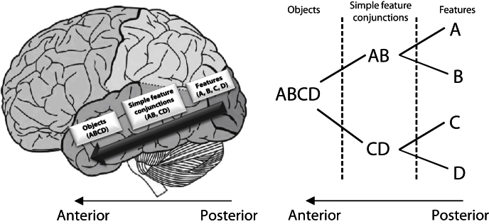
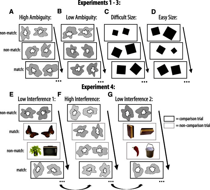
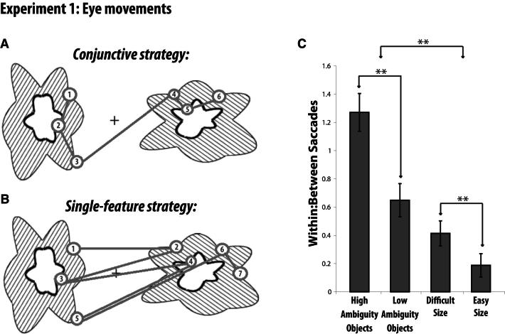
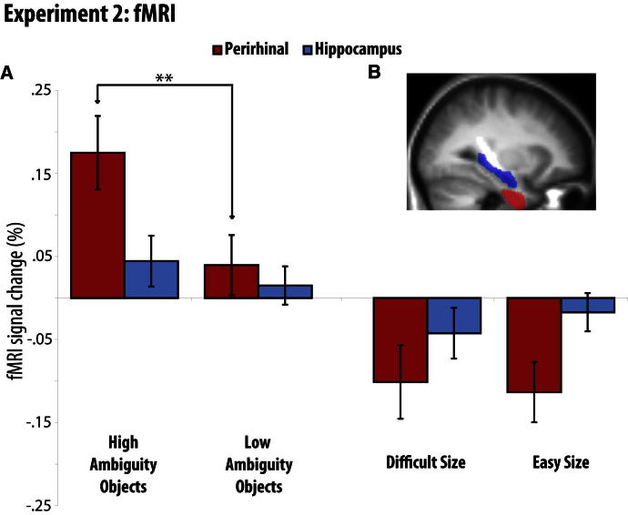
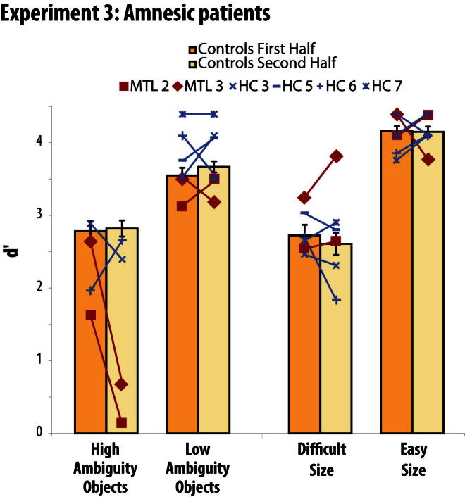
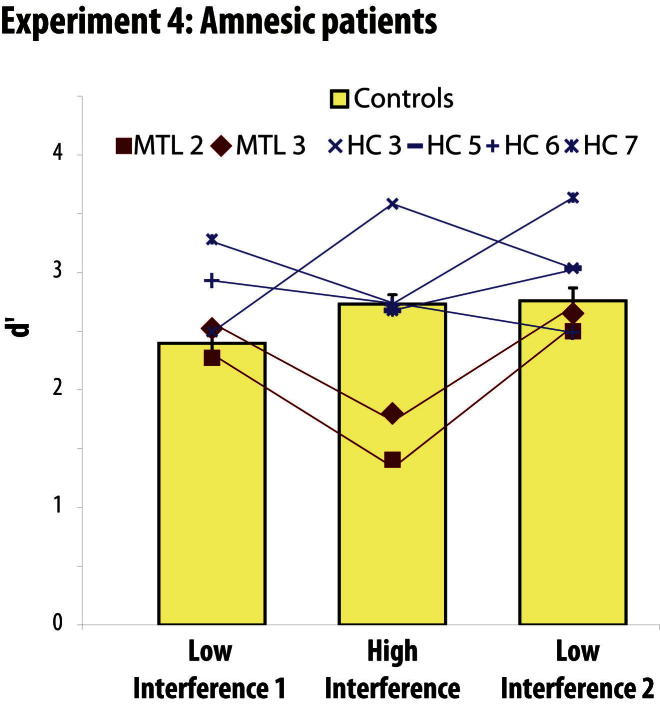
Memory and perception have long been considered separate cognitive processes, and amnesia resulting from medial temporal lobe (MTL) damage is thought to reflect damage to a dedicated memory system. Recent work has questioned these views, suggesting that amnesia can result from impoverished perceptual representations in the MTL, causing an increased susceptibility to interference. Using a perceptual matching task for which fMRI implicated a specific MTL structure, the perirhinal cortex, we show that amnesics with MTL damage including the perirhinal cortex, but not those with damage limited to the hippocampus, were vulnerable to object-based perceptual interference. Importantly, when we controlled such interference, their performance recovered to normal levels. These findings challenge prevailing conceptions of amnesia, suggesting that effects of damage to specific MTL regions are better understood not in terms of damage to a dedicated declarative memory system, but in terms of impoverished representations of the stimuli those regions maintain.


Figures

Figure 1
Figure 1
Representational-Hierarchical Theory (A) Lateral view of the human cerebral cortex demonstrating the ventral visual stream (VVS) object processing pathway according to the representational-hierarchy theory (Cowell et al., 2010a). The perirhinal cortex is proposed to reside at the apex of this processing pathway, containing complex representations of objects. (B) The proposed organization of visual object representations in the VVS. A, B, C, and D refer to relatively simple object features represented in posterior regions. More complex conjunctions of these features are stored in more anterior regions, including perirhinal cortex. Figure adapted from Bussey and Saksida (Bussey and Saksida, 2002).
Figure 2
Figure 2
Visual Discrimination Task Participants indicated whether two simultaneously presented stimuli were a match or a non-match. For experiments 1–3, there were four conditions: (A) High Ambiguity Objects, (B) Low Ambiguity Objects, (C) Difficult Size, (D) Easy Size. The objects were defined by three features: inner shape, outer shape, and fill pattern. For High Ambiguity nonmatch trials, only one of these three features differed, whereas for Low Ambiguity nonmatch trials, all three features differed. Thus, the High Ambiguity Object condition placed a greater demand on high-level conjunctive representations and analysis of the object as a whole, which was confirmed by an analysis of eye movement patterns (see Experiment 1). In the Size control task, participants decided if two rotated squares were the same size. (E–G) Example stimuli and trial order from the Low and High Interference conditions in experiment 4. For the Low Interference condition, a High Ambiguity Object trial was always followed by two trials involving perceptually distinct, colored objects (30 High Ambiguity Object trials in total). The High Interference condition was a straight block of 88 consecutive High Ambiguity Object trials. To avoid confounding effects of fatigue, the order of testing conditions was: Low Interference 1, High Interference, Low Interference 2. We compared performance on every third trial only (black boxes), thus ensuring that for each condition our comparison trials were 30 High Ambiguity Object trials with matched stimulus schedules. Across all experiments, all objects were trial unique, though the individual features (e.g., shape segments, fill patterns) repeated across trials.
Figure 3
Figure 3
Experiment 1 Example of idealized viewing patterns associated with (A) viewing the stimulus as a whole object (conjunctive strategy) and (B) viewing the stimulus as a series of individual features (single-feature strategy). Each fixation is shown by a numbered circle indicating the order of the fixation; gray lines connecting the fixations indicate saccades. (C) Fixation patterns across the four conditions in experiment 1. The critical ratio of saccades within an item relative to saccades between items indicated that the High Ambiguity Object condition was associated with a greater degree of conjunctive processing. The individual within-item and between-item saccade averages that comprise this ratio are shown in Figure S1. Error bars represent SEM; ∗∗p < 0.001.
Figure 4
Figure 4
Experiment 2 (A) Percent BOLD change relative to mean over all voxels and scans, mean-corrected over conditions, within the PRC and hippocampal anatomical regions of interest (images were not smoothed). Activity in the PRC was modulated by the degree of feature ambiguity, but not general task difficulty. Activity in the hippocampus was not sensitive to either feature ambiguity or control task difficulty. Error bars represent SEM of the difference between each condition and its relevant control (i.e., High Ambiguity Objects – Low Ambiguity Objects or Difficult Size – Easy Size), ∗∗p < 0.001. Accuracy and reaction time data are reported in the Supplemental Information (Table S1). In brief, reduced accuracy was found in High relative to Low Ambiguity conditions, and Difficult relative to Easy control conditions, as expected. Importantly, the planned comparison revealed no greater difference in accuracy between High Ambiguity and Low Ambiguity Objects than between Difficult and Easy Size (t(19) = 0.16), suggesting that fMRI effects of feature ambiguity are not confounded by difficulty. (B) Critical regions of interest superimposed on the mean structural image across participants (PRC in red, hippocampal in blue). See also Figures S2 and S3.
Figure 5
Figure 5
Experiment 3 d′ scores for each individual patient and the mean of their controls, split according to the first and second half of each condition in experiment 3 (patients performed 72 consecutive trials of each condition, with condition order counterbalanced). There was a dramatic decline in performance of patients whose lesion included PRC (MTL cases) as the High Ambiguity condition progressed. This performance decline was limited to the MTL cases on the High Ambiguity Object condition: it was not observed on any other condition or in any other participant group. Moreover, the MTL cases performed normally on the equally challenging Difficult Size condition. Cases with hippocampal lesions (HC cases) performed normally on all conditions. Error bars represent SEM. The separate control groups (age-matched to either the MTL or HC individuals) showed no evidence of differing, in terms of overall performance or relative performance across conditions (all F < 0.7, p > 0.6), and thus are plotted as a single group. A paired t test showed no evidence that control participants found the High Ambiguity Object condition more difficult than the Difficult Size control condition (t(21) = 0.5, p = 0.6), suggesting that the deficit in the MTL cases was not driven by task difficulty. See also Figure S4.
Figure 6
Figure 6
Experiment 4 d′ scores for each individual patient and the mean of their controls in experiment 4. Patients whose lesions included PRC (MTL cases) were impaired on the High Interference condition, but their performance was rescued by reducing the degree of interference. Cases with hippocampal lesions (HC cases) performed normally on all conditions. Error bars represent SEM. The separate control groups showed no evidence of differing, in terms of overall performance or relative performance across conditions (all F < 1, p > 0.4), and thus are plotted as a single group. See also Figure S4.

Comment in

Similar articles

Cited by

References

    1. Barense M.D., Bussey T.J., Lee A.C., Rogers T.T., Davies R.R., Saksida L.M., Murray E.A., Graham K.S. Functional specialization in the human medial temporal lobe. J. Neurosci. 2005;25:10239–10246. - PMC - PubMed
    1. Barense M.D., Gaffan D., Graham K.S. The human medial temporal lobe processes online representations of complex objects. Neuropsychologia. 2007;45:2963–2974. - PubMed
    1. Barense M.D., Henson R.N.A., Lee A.C., Graham K.S. Medial temporal lobe activity during complex discrimination of faces, objects, and scenes: Effects of viewpoint. Hippocampus. 2010;20:389–401. - PMC - PubMed
    1. Barense M.D., Rogers T.T., Bussey T.J., Saksida L.M., Graham K.S. Influence of conceptual knowledge on visual object discrimination: insights from semantic dementia and MTL amnesia. Cereb. Cortex. 2010;20:2568–2582. - PubMed
    1. Barense M.D., Henson R.N., Graham K.S. Perception and conception: temporal lobe activity during complex discriminations of familiar and novel faces and objects. J. Cogn. Neurosci. 2011;23:3052–3067. - PubMed

Publication types