. 2012 Jul 13:10:30.
doi: 10.1186/1477-3155-10-30.

Chimeric Hepatitis B core antigen virus-like particles displaying the envelope domain III of dengue virus type 2

Affiliations

Chimeric Hepatitis B core antigen virus-like particles displaying the envelope domain III of dengue virus type 2

Upasana Arora et al. J Nanobiotechnology. .

Abstract

Background: Dengue is a global public health problem for which no drug or vaccine is available. Currently, there is increasing interest in developing non-replicating dengue vaccines based on a discrete antigenic domain of the major structural protein of dengue viruses (DENVs), known as envelope domain III (EDIII). The use of bio-nanoparticles consisting of recombinant viral structural polypeptides, better known as virus-like particles (VLPs), has emerged as a potential platform technology for vaccine development. This work explores the feasibility of developing nanoparticles based on E. coli-expressed recombinant Hepatitis B virus core antigen (HBcAg) designed to display EDIII moiety of DENV on the surface.

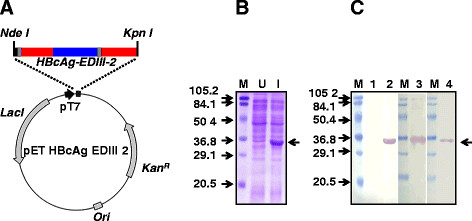
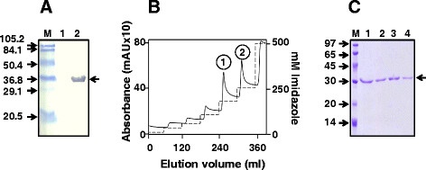
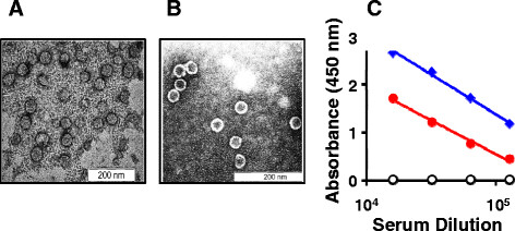
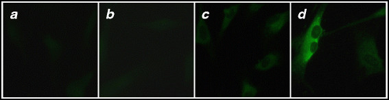
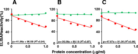
Findings: We designed a synthetic gene construct encoding HBcAg containing an EDIII insert in its c/e1 loop. The fusion antigen HBcAg-EDIII-2 was expressed in E. coli, purified to near homogeneity using Ni+2 affinity chromatography and demonstrated to assemble into discrete 35-40 nm VLPs by electron microscopy. Competitive ELISA analyses showed that the EDIII-2 moieties of the VLPs are accessible to anti-EDIII-2-specific monoclonal and polyclonal antibodies, suggesting that they are surface-displayed. The VLPs were highly immunogenic eliciting high titer anti-EDIII-2 antibodies that were able to recognize, bind and neutralize infectious DENV based on ELISA, immunofluorescence and virus-neutralization assays.

Conclusion: This work demonstrates that HBcAg-derived nanoparticles can serve as a useful platform for the display of DENV EDIII. The EDIII-displaying nanoparticles may have potential applications in diagnostics/vaccines for dengue.


Figures

Figure 1
Figure 1
Design and expression of HBcAg-EDIII-2 antigen inE. coli. (A) A map of the HBcAg-EDIII-2 expression vector. The synthetic HBcAg-EDIII-2 gene is inserted under the control of the phage T7 promoter (pT7) in pET29a. The organization of different segments of this fusion gene is indicated in colour as follows. The HBcAg- and EDIII-2-encoding regions are shown in red and blue, respectively. The 6x His tag-encoding sequences at the 5’end is shown in black. The two linker-encoding sequences, the first following the 6x His tag and the second after the EDIII-2 encoding sequences, are shown in grey. Other abbreviations are as follows. Lac I: Lac repressor gene; KanR: Kanamycin marker; Ori: Replication origin sequences. (B) SDS-PAGE analysis of recombinant HBcAg-EDIII-2 expression. This panel displays the Coomassie-stained polypeptide profiles of lysates prepared from un-induced (U) and induced (I) E. coli cells harboring the plasmid shown in A. Pre-stained protein molecular weight markers were run in lane ‘M’. Their sizes (in kDa) are shown at the left of the panel. The arrow on the right indicates the position of the recombinant HBcAg-EDIII-2 protein. (C) Immunoblot analyses of recombinant protein expression. Aliquots of un-induced and induced cell lysates (described in panel ‘B’) were electrophoresced, electroblotted onto nitrocellulose membranes and probed with anti-EDIII mAb 24A12 (lane 2), penta His mAb (lane 3), or anti-HBcAg mAb ab8638 (lane 4). An aliquot of the un-induced cell lysate was probed with mAb 24A12 (lane 1). Pre-stained protein size markers were run in lanes marked ‘M’. Their sizes (in kDa) are indicated to the left of the first blot. The arrow to the right indicates the position of the recombinant HBcAg-EDIII-2 protein.
Figure 2
Figure 2
Affinity purification of the recombinant HBcAg-EDIII-2 protein under denaturing conditions. (A) Western blot analysis of localization of HBcAg-EDIII-2 expression. Induced cells were sonicated and centrifuged. The resultant supernatant (lane 1) and pellet (lane 2) fractions were boiled in Laemmli loading buffer, electrophoresced under denaturing conditions and subjected to immunoblot analysis using mAb24A12 to identify the recombinant HBcAg-EDIII-2 protein. (B) Ni2+-affinity purification of HBcAg-EDIII-2 from induced E. coli cells. The insoluble pellet obtained after sonication of induced cells was purified using Ni2+-Sepharose under denaturing conditions. The solid curve represents the chromatographic profile obtained by measurement of absorbance at 280 nm. The two peaks discernible in the elution profile are numbered 1 and 2. The dotted curve represents the imidazole step-gradient employed for elution. (C) SDS-PAGE analysis of fractions corresponding to peaks 1 (lanes 1 & 2) and 2 (lanes 3 & 4) shown in panel ‘B’. Low molecular weight protein markers were run in lane ‘M’; their sizes (in kDa) are indicated to the left of the panel. The arrow at the right of the panels A and C indicates the position of the HBcAg-EDIII-2 protein.
Figure 3
Figure 3
Characterization of the purified HBcAg-EDIII-2 antigen. (A) The panel depicts VLPs formed by purified HBcAg-EDIII-2 protein (expressed in P.pastoris). Peaks 1 and 2 (shown in Figure 2B) were pooled, dialyzed, stained with 1% uranyl acetate and visualized under the electron microscope. (B) Electron microscopic visualization of the purified HBcAg carrier (expressed in E. coli). (C) Evaluation of the immunogenicity of the HBcAg-EDIII-2 antigen by ELISA. Antisera from mice immunized with the fusion antigen (solid red circles) and its precursors (HBcAg: empty black circles; EDIII-2: solid blue diamonds) were analyzed in an ELISA using recombinant EDIII-2 antigen (produced using P. pastoris) as the coating antigen.
Figure 4
Figure 4
Evaluation of antibodies elicited by HBcAg-EDIII-2 fusion antigen by indirect immunofluorescence assay. Sera from mice that were mock-immunized (panel a), and immunized with HBcAg (panel b), EDIII-2 (panel c) or HBcAg-EDIII-2 (panel d) antigens were used as the primary antibody to pick up DENV-2 in infected BHK-21 cells, in conjunction with an anti-mouse IgG-FITC conjugate.
Figure 5
Figure 5
Competitive ELISA. Anti-EDIII mAb24A12 (panel A), anti-EDIII-T antiserum (panel B) and anti-HBcAg-EDIII-2 antiserum (panel C) were separately pre-incubated with varying concentrations of purified HBcAg (green curves) and HBcAg-EDIII-2 (red curves) VLPs, and analyzed for residual anti-EDIII-2 antibodies using purified P. pastoris-expressed EDIII-2 antigen as the coating antigen. ELISA reactivity in the absence of any added protein in the pre-incubation step was taken to represent 100%. The regression equations for the HBcAg-EDIII-2 competition assays (red curves) are shown just above the horizontal axis in each panel.

Similar articles

Cited by

References

    1. World Health Organization Factsheet No117. Dengue and dengue haemorrhagic fever. , ; 2012. www.who.int/mediacentre/factsheets/fs117/en/[Accessed April 27, 2012]
    1. Gubler DJ, Kuno G, Markoff L. In: Fields Virology. 5. Knipe DM, Howley PM, editor. Wolters Kluwer and Lippincott Williams & Wilkins, Philadelphia; 2007. Flaviviruses; pp. 1153–1252.
    1. Swaminathan S, Batra G, Khanna N. Dengue vaccines: state of the art. Expert Opin Ther Patents. 2010;20:819–835. doi: 10.1517/13543771003767476. - DOI - PubMed
    1. Schmitz J, Roehrig J, Barrett A, Hombach J. Next generation dengue vaccines: a review of candidates in preclinical development. Vaccine. 2011;29:7276–7284. doi: 10.1016/j.vaccine.2011.07.017. - DOI - PubMed
    1. Guzman MG, Hermida L, Bernardo L, Ramirez R, Guillén G. Domain III of the envelope protein as a dengue vaccine target. Expert Rev Vaccines. 2010;9:137–147. doi: 10.1586/erv.09.139. - DOI - PubMed

Publication types

MeSH terms