. 2012 Jul 13;91(1):109-21.
doi: 10.1016/j.ajhg.2012.05.026.

Metabolic correction of congenital erythropoietic porphyria with iPSCs free of reprogramming factors

Affiliations

Metabolic correction of congenital erythropoietic porphyria with iPSCs free of reprogramming factors

Aurélie Bedel et al. Am J Hum Genet. .

Abstract

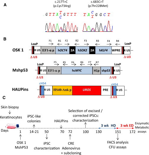
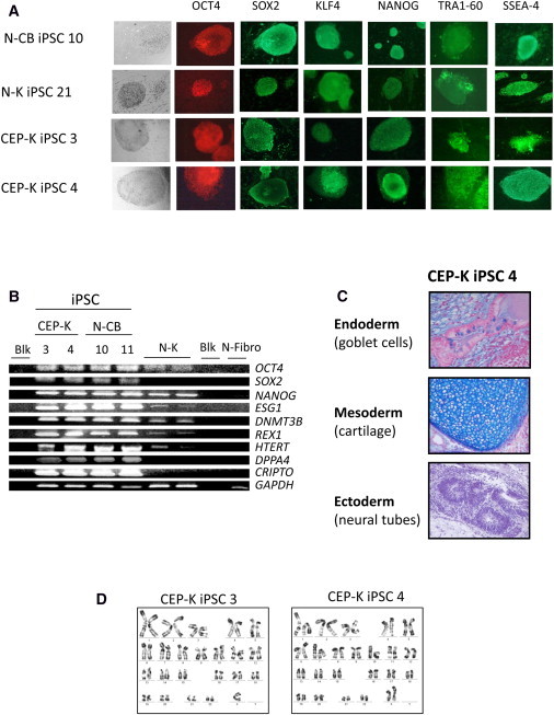
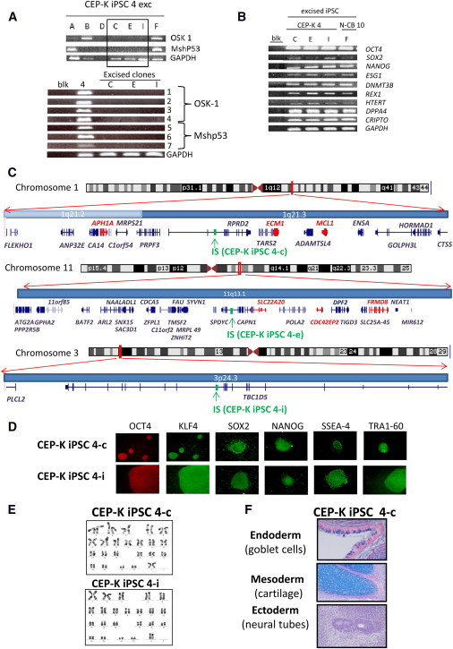
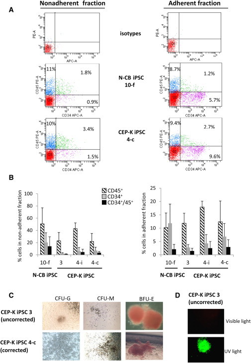
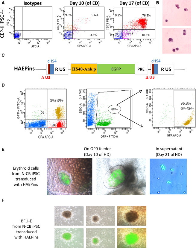
Congenital erythropoietic porphyria (CEP) is due to a deficiency in the enzymatic activity of uroporphyrinogen III synthase (UROS); such a deficiency leads to porphyrin accumulation and results in skin lesions and hemolytic anemia. CEP is a candidate for retrolentivirus-mediated gene therapy, but recent reports of insertional leukemogenesis underscore the need for safer methods. The discovery of induced pluripotent stem cells (iPSCs) has opened up new horizons in gene therapy because it might overcome the difficulty of obtaining sufficient amounts of autologous hematopoietic stem cells for transplantation and the risk of genotoxicity. In this study, we isolated keratinocytes from a CEP-affected individual and generated iPSCs with two excisable lentiviral vectors. Gene correction of CEP-derived iPSCs was obtained by lentiviral transduction of a therapeutic vector containing UROS cDNA under the control of an erythroid-specific promoter shielded by insulators. One iPSC clone, free of reprogramming genes, was obtained with a single proviral integration of the therapeutic vector in a genomic safe region. Metabolic correction of erythroblasts derived from iPSC clones was demonstrated by the disappearance of fluorocytes. This study reports the feasibility of porphyria gene therapy with the use of iPSCs.


Figures

Figure 1
Figure 1
CEP Characterization and Schematic Drawing of iPSC Generation (A) Genotyping of keratinocytes from a CEP-affected individual compound heterozygous for UROS mutations c.217T>C (p.Cys73Arg) and c.683C>T (p.Thr228Met). (B) Schematic representation of the proviral form of the lentivectors used. OSK 1 is an excisable single polycistronic vector coexpressing OCT4, SOX2, and KLF4 cDNAs linked with the porcine teschovirus-1 2A sequence. Mshp53 coexpresses MYC and a shRNA against TP53; both vectors are flanked by loxP sites. HAUPins contains the UROS cDNA under the control of the erythroid chimeric HS-40 enhancer/ankyrin promoter. Arrows show the position of forward (F) and reverse (R) primers used for proviral integration analyses. The following abbreviations are used: HS40, enhancer of HBA1 (MIM 141800); Ank p, ankyrin promoter of ANKYRIN-1 (MIM 612641); UROS, cDNA of uroporphyrinogene III synthase; H1p, mammalian polymerase III H1 promoter; EF1α-p, elongation factor-1 alpha promoter; WPRE, woodchuck posttranscriptional regulatory element; PRE, a mutated WPRE sequence (WPREmut6); and cHS4, element of the chicken β-globin hypersensitivity site 4. (C) Schematic strategy used for reprogramming human keratinocytes from a CEP-affected person and for HSC differentiation. The following abbreviations are used: HD, hematopoietic differentiation; and ED, erythroid differentiation.
Figure 2
Figure 2
Characterization of iPSC Clones (A) Representative immunofluorescence of pluripotency markers in human iPSC clones derived from normal CD34+ CB cells (N-CB iPSC 10) and in keratinocytes derived from a normal individual (N-K iPSC 21) and a CEP-affected individual (CEP-K iPSC 3 and CEP-K iPSC 4); staining is with anti-OCT4, anti-SOX2, anti-KLF4, anti-NANOG, anti-SSEA-4, and anti-TRA1-60. MEFs surrounding human iPSCs served as a negative control for immunofluorescence (magnification ×100 or ×200). (B) Expression of pluripotency-associated genes (OCT4, SOX2, NANOG, ESG1, DNMT3B, REX1, HTERT, DPPA4, and CRIPTO) by RT-PCR cycles (28) from two independent CEP-K iPSC clones and two N-CB iPSC clones. Primary N-K cells and primary normal human fibroblast (N-Fibro) cells were used as controls. GAPDH served as an internal positive control. The following abbreviation is used: Blk, blank (PCR performed without cDNA). (C) Alcian-blue staining of histological sections of a representative teratoma derived from human CEP-K iPSC 4 shows tissues of all three germ layers (magnification ×200). (D) Representative karyotypic analysis of two human CEP-K iPSC clones (CEP-K iPSC 3 and CEP-K iPSC 4).
Figure 3
Figure 3
Characterization of iPSC Clones Free of Reprogramming Transgene (A) Upper panel: PCRs for the integrated vectors OSK 1 and Mshp53 in seven CEP-K iPSC 4 subclones pretreated by CRE adenovirus (subclones are A, B, C, D, E, F, and I). Lower panel: Multiple PCRs performed on DNA from the three excised clones (C, E, and I) and the nonexcised iPSC 4 with seven couples of primers described in (B) for detecting the presence of portions of the recombinant provirus. (B) Expression of pluripotency-associated genes by RT-PCR from three independent iPSC subclones from CEP-K iPSC 4 and one iPSC subclone from N-CB iPSC 10. (C) Chromosome ideograms and graphics depicting 300 kb of the human genome on both sides of the proviral IS (in green) of HAUPins from CEP-K iPSC 4-c (upper panel), from CEP-K iPSC 4-e (middle panel), and from CEP-K iPSC 4-i (lower panel). Graphics were obtained with the UCSC Genome Graphs tool. All known genes present in the genomic region are shown (genes implicated in cancer according to the allOnco database are shown in red). (D) Representative immunofluorescence of pluripotency markers in human iPSC clones that are free of reprogramming transgene and derived from CEP-K iPSC 4-c and 4-i (magnification ×100 or ×200). (E) Karyotypic analysis of the two excised clones (4-c and 4-i). (F) Alcian-blue staining of histological sections of a teratoma derived from human CEP-K iPSC 4-c, free of reprogramming transgene, shows tissues of all three germ layers.
Figure 4
Figure 4
Efficient Hematopoietic Differentiation of iPSCs (A) Representative FACS analysis of CD45+ and CD34+ cells obtained from N-CB iPSC 10-f (upper panels) and CEP-K iPSC 4-c (lower panels), both free of reprogramming transgenes, after hematopoietic differentiation (at day 21) in nonadherent fraction (left panels) and in adherent fraction (right panels) from the same experiment. (B) Bar graphs show the average percentages of CD45+, CD34+, and CD34+ and CD45+ cells obtained from four iPSC clones (N-CB iPSC 10-f, uncorrected CEP-K iPSC 3, and corrected CEP-K iPSCs 4-i and 4-c) in nonadherent fractions (left panel) and in adherent fractions (right panel) at day 21 of hematopoietic differentiation (n = 5 independent experiments, mean ± SD). No statistical difference was observed between clones. (C) Bright-field microscopy of CFUs (a granulocytic CFU [CFU-G], a monocytic CFU [CFU-M], and an erythroid burst-forming unit [BFU-E]) in methylcellulose medium by hematopoietic cells obtained from uncorrected (upper panel) or corrected (lower panel) CEP-K iPSCs (magnification ×100). (D) Inverted microscopy of a BFU-E in methylcellulose derived from the uncorrected CEP-K iPSC 3 under visible (upper panel) or UV light (lower panel).
Figure 5
Figure 5
Efficient Erythroid Differentiation of HSCs Derived from iPSCs with Specific Transgene Expression (A) Representative flow-cytometry data of in vitro erythroid-differentiation (ED) cultures from CEP-K iPSC 4-i. Cells were stained at day 10 and at day 17 of ED with anti-CD71-phycoerythrin (PE)- and anti-GPA-APC-conjugated antibodies (or they were stained with control isotype antibodies conjugated with PE and APC; left panels). (B) Erythroblast maturation was evaluated at day 24 of ED by staining with May-Grünwald-Giemsa. Photographs show the late stage of erythroid maturation. (C) Schema of the proviral form of the HAEPins control vector containing EGFP under the control of the HS40/ankyrin erythroid promoter. (D) Representative FACS analysis of hematopoietic progenitors at day 21 of hematopoietic differentiation (HD) from the N-CB iPSCs previously transduced with the HAEPins lentivector. (E) At day 10 of HD, formation of early erythroblast cells was revealed by GFP expression in sacks on OP9 stroma (middle and left panels) and by erythroblast cell suspension at day 21 of HD (right panel). (F) Inverted microscopy of BFU-E colonies under a visible-light microscope (upper panels) and merged with GFP expression detected under UV light (lower panels).
Figure 6
Figure 6
Enzymatic and Metabolic Correction (A) UROS enzymatic activities in primary N-K and deficient CEP-K cells and in erythroid cells derived from N-CB iPSC 10-f, CEP-K iPSC 3 (uncorrected), and CEP-K iPSC 4-i (corrected) clones. (B) Bar graphs show average percentages of fluorocytes gated on erythroid GPA+ cells from three independent experiments (mean ± SD). (C) Representative FACS analysis of the fluorocytes gated on erythroid GPA+ cells derived from N-CB iPSC 10-f and uncorrected CEP-K iPSC 3 (upper panels) and from two corrected clones (4-i and 4-c; lower panels) at day 21 of erythroid differentiation.

Similar articles

Cited by

References

    1. Anderson K.E., Sassa S., Bishop D.F., Desnick R.J. Disorders of heme biosynthesis: X-linked sideroblastic anemia and the porphyrias. In: Scriver R., Beaudet A.L., Sly W.S., Valle E., editors. The Metabolic and Molecular Bases of Inherited Disease. C McGraw-Hill; New York: 2001. pp. 2961–3062.
    1. Richard E., Robert-Richard E., Ged C., Moreau-Gaudry F., de Verneuil H. Erythropoietic porphyrias: animal models and update in gene-based therapies. Curr. Gene Ther. 2008;8:176–186. - PubMed
    1. Shaw P.H., Mancini A.J., McConnell J.P., Brown D., Kletzel M. Treatment of congenital erythropoietic porphyria in children by allogeneic stem cell transplantation: A case report and review of the literature. Bone Marrow Transplant. 2001;27:101–105. - PubMed
    1. Kauffman L., Evans D.I.K., Stevens R.F., Weinkove C. Bone-marrow transplantation for congenital erythropoietic porphyria. Lancet. 1991;337:1510–1511. - PubMed
    1. Lagarde C., Hamel-Teillac D., De Prost Y., Blanche S., Thomas C., Fischer A., Nordmann Y., Ged C., De Verneuil H. [Allogeneic bone marrow transplantation in congenital erythropoietic porphyria. Gunther's disease] Ann. Dermatol. Venereol. 1998;125:114–117. - PubMed

Publication types

MeSH terms

Substances