. 2013 Apr;131(4):1117-29, 1129.e1-5.
doi: 10.1016/j.jaci.2012.05.036. Epub 2012 Jul 11.

Patients with cystic fibrosis have inducible IL-17+IL-22+ memory cells in lung draining lymph nodes

Affiliations

Patients with cystic fibrosis have inducible IL-17+IL-22+ memory cells in lung draining lymph nodes

Yvonne R Chan et al. J Allergy Clin Immunol. 2013 Apr.

Abstract

Background: IL-17 is an important cytokine signature of the TH differentiation pathway TH17. This T-cell subset is crucial in mediating autoimmune disease or antimicrobial immunity in animal models, but its presence and role in human disease remain to be completely characterized.

Objective: We set out to determine the frequency of TH17 cells in patients with cystic fibrosis (CF), a disease in which there is recurrent infection with known pathogens.

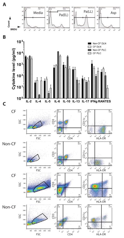
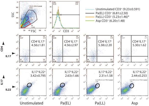
Methods: Explanted lungs from patients undergoing transplantation or organ donors (CF samples=18; non-CF, nonbronchiectatic samples=10) were collected. Hilar nodes and parenchymal lung tissue were processed and examined for TH17 signature by using immunofluorescence and quantitative real-time PCR. T cells were isolated and stimulated with antigens from Pseudomonas aeruginosa and Aspergillus species. Cytokine profiles and staining with flow cytometry were used to assess the reactivity of these cells to antigen stimulation.

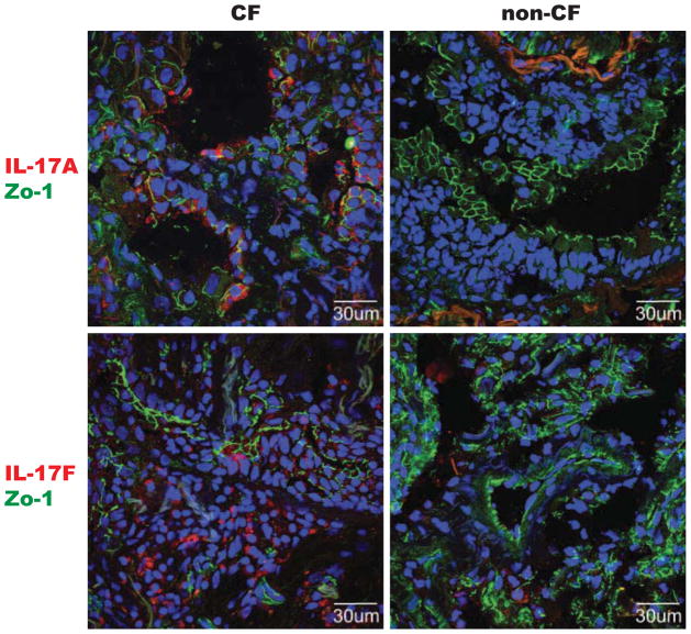
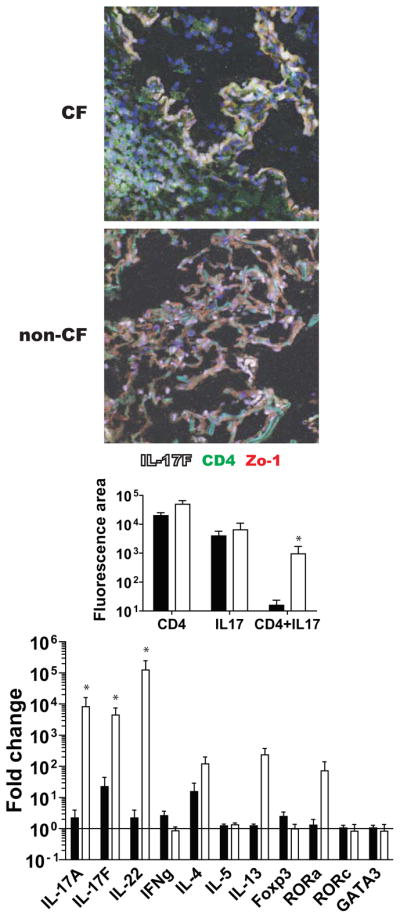
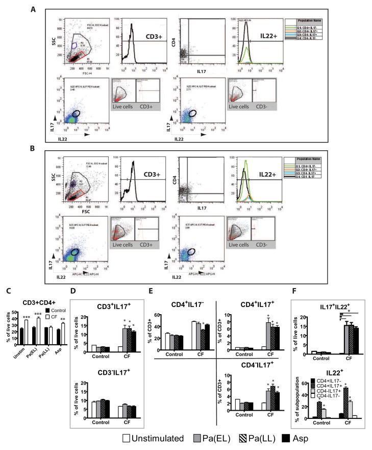
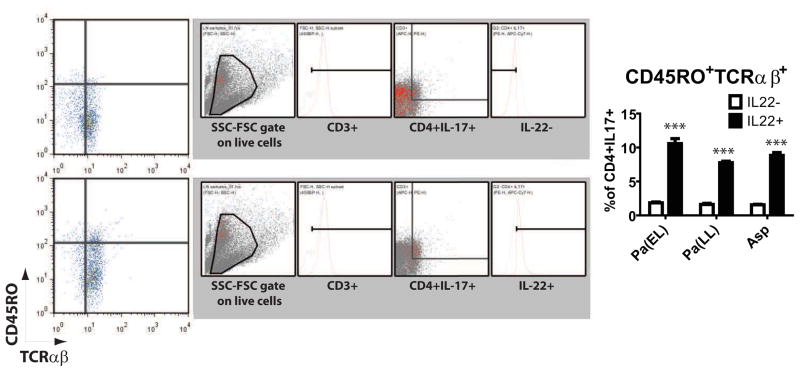
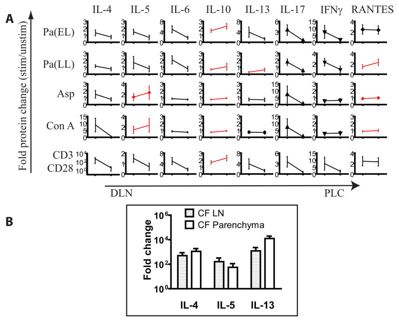
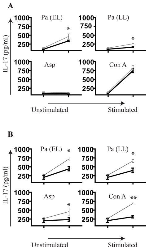
Results: We found a strong IL-17 phenotype in patients with CF compared with that seen in control subjects without CF. Within this tissue, we found pathogenic antigen-responsive CD4+IL-17+ cells. There were double-positive IL-17+IL-22+ cells [TH17(22)], and the IL-22+ population had a higher proportion of memory characteristics. Antigen-specific TH17 responses were stronger in the draining lymph nodes compared with those seen in matched parenchymal lungs.

Conclusion: Inducible proliferation of TH17(22) with memory cell characteristics is seen in the lungs of patients with CF. The function of these individual subpopulations will require further study regarding their development. T cells are likely not the exclusive producers of IL-17 and IL-22, and this will require further characterization.


Figures

Figure 1
Figure 1. IL-17 positive cells are prominent in CF lung tissue
Parenchymal lung tissue stained for IL-17A (red, top two) or IL-17F (red, bottom two) and co-stained with tight junction protein-1 (Zo-1, green, all) to delineate anatomic structure. DAPI counterstaining (blue) was done to highlight nuclei. Representative images from CF patients (left two) and non-CF controls (right two, patients transplanted for primary pulmonary hypertension and idiopathic pulmonary fibrosis) are shown.
Figure 2
Figure 2. CF lungs demonstrate higher numbers of dual positive CD4+IL-17+ cells and have stronger Th17 cytokine message profile
(Top photomicrographs) Representative images of parenchymal lung tissue stained for IL-17F (white) and co-stained with anti CD4 (green) and Zo-1 (red). (Middle graph) Quantitative confocal analysis of all image files obtained, compared by genotype control ■ (n = 9) vs CF □ (n = 12). (Bottom graph) qPCR reveals increased Th17 transcripts in CF lungs. ΔΔCt values for control ■ (n = 9) and CF □ (n = 12) are shown. IL-17A, IL-17F and IL-22 are upregulated in CF lung tissue while related transcript RORa also trends higher in CF. * = p < 0.05
Figure 3
Figure 3. Antigen stimulation preparations and baseline characteristics of CF and non-CF samples
(A) CF DLN cells were stimulated as described and BRDU histograms from antigen concentrations that were chosen are shown. (B) Cultured DLN and PLC supernatants were assayed by multiplex cytokine bead array and IL-17 mean absolute values with SEM for each cohort and tissue sample are graphed. IL-17 levels are not remarkably different among all CF (n = 10) or control (n = 4) samples prior to stimulation. Culture media included IL-2. (C) DLN (top two rows of plots) and PLC (bottom two rows of plots) are assessed prior to culture with antigen stimulation. Representative dot plots are shown. Total events examined by size (FSC) vs granularity (SSC) were first plotted and a live cell gate was drawn. This live cell gate was subsequently analyzed for CD3 vs CD4 and CD11c vs HLA-DR.
Figure 4
Figure 4. IL-17+ and IL-22+ T cells are increased in CF
Representative gating strategy for non-CF (A) and CF (B) is shown on a Pa(EL) stimulated sample. Total events were analyzed by size (FSC) vs granularity (SSC) and a live cell gate (largest, gray gate) was drawn. This gate was further examined by univariate histogram analysis of CD3 status (top row, second from left). Gated CD3+ cells were then analyzed by CD4 vs IL-17 (top row, third from left) and proportion of IL-22+ cells in each quadrant graphed in the second histogram. Second row in each lettered section shows all live cells that were CD3+ (first bivariate plot) and CD3− (second bivariate plot) analyzed for IL-17 vs IL-22. An IL-17+IL-22+ double positive gate was drawn and backgating to show ancestry is shown in the gray inset. (C) Non-CF ■ (n = 4) and CF □ (n = 7) population results of bivariate analysis of the live cell gate by CD3 vs CD4. (D) Cohort statistics by antigen stimulation of CD3+ and CD3− cells that are IL-17+. (E) Breakdown of quadrant statistics in the CD4 vs IL-17 plot by genotype and antigen stimulation. (F, top) Cohort statistics of the IL-17+IL-22+ double positive gate by genotype and antigen stimulation. (F, bottom) IL-22+ statistics for each quadrant in the CD4 vs IL-17 analysis for each genotype and antigen stimulation. Figure legend for D, E, F is shown on bottom unless otherwise noted in F, bottom graph. * p < 0.05; ** p < 0.001; *** p < 0.0001.
Figure 5
Figure 5. IL-22+ status correlates with memory T cell characteristics
IL-22+ and IL22− cells in the live cell gate were analyzed for CD45RO vs TCRαβ expression. Representative plots with backgating and ancestry shown in the gray inset next to each bivariate plot for Pa(EL) stimulated CF DLN. IL-22 positivity in CD4+ IL-17+ cells correlated to CD45RO+TCRab+ status (right graph) * p < 0.05; ** p < 0.001; *** p < 0.0001.
Figure 6
Figure 6. PLC shows minimal to modest IL-17 and IL-22 response to antigen stimulation
Representative plots on a CF PLC sample are shown. (Top row) Live cell gate in SSC vs FSC plot was examined for CD3 status (histogram). Statistics for each stimulation are shown in the histogram legend. One stimulation condition resulted in statistically significant reduction in the number of CD3+ cells relative to the unstimulated sample [Pa(LL)]. The live cell gate was then examined for CD4 vs IL-17 (second row of plots) with statistics for the CD4+IL-17+ quadrant shown for each antigen stimulation. Similarly the live cell gate was examined for IL-17 vs IL-22 in each antigen stimulation and statistics are shown for the IL-17+IL-22+ quadrant (bottom row of plots). * p < 0.05
Figure 7
Figure 7. Presence of Th2 or Treg cytokines in the parenchyma may blunt IL-17 response there
(A) Average normalized antigen stimulated cytokine values for CF DLN and CF PLC were juxtaposed. Cytokines in which PLC > DLN are in red. Among cytokines exhibiting an increase in PLC relative to DLN are IL-10, IL-13 and RANTES for Pa stimulation; IL-5, IL-10 and IL-13 for Asp. (B) qPCR of Th2 cytokines in CF lymph node and parenchyma show a trend toward higher signals in the parenchyma.
Figure 8
Figure 8. Neutralization of Th1 and Th2 cytokines augments the IL-17 response
(A) CF DLN (n = 3) were cultured with indicated antigen along with neutralizing antibodies to IL-4 and IFNγ together ( formula image) and compared to no neutralization (—) with 20 μg/ml of IgG. (B) IL-13 and IFNγ were also neutralized (n = 3).. * p < 0.05, ** p < 0.001

Similar articles

Cited by

References

    1. Mosmann TR, Cherwinski H, Bond MW, Giedlin MA, Coffman RL. Two types of murine helper T cell clone. I. Definition according to profiles of lymphokine activities and secreted proteins. J Immunol. 1986;136:2348–57. - PubMed
    1. Garvy BA, Wiley JA, Gigliotti F, Harmsen AG. Protection against Pneumocystis carinii pneumonia by antibodies generated from either T helper 1 or T helper 2 responses. Infect Immun. 1997;65:5052–6. - PMC - PubMed
    1. Yao Z, Fanslow WC, Seldin MF, Rousseau AM, Painter SL, Comeau MR, et al. Herpesvirus Saimiri encodes a new cytokine, IL-17, which binds to a novel cytokine receptor. Immunity. 1995;3:811–21. - PubMed
    1. Yao Z, Painter SL, Fanslow WC, Ulrich D, Macduff BM, Spriggs MK, et al. Human IL-17: a novel cytokine derived from T cells. J Immunol. 1995;155:5483–6. - PubMed
    1. Rouvier E, Luciani MF, Mattei MG, Denizot F, Golstein P. CTLA-8, cloned from an activated T cell, bearing AU-rich messenger RNA instability sequences, and homologous to a herpesvirus saimiri gene. J Immunol. 1993;150:5445–56. - PubMed

Publication types

MeSH terms