

Movement- and behavioral state-dependent activity of pontine reticulospinal neurons
- PMID: 22796072
- PMCID: PMC3424299
- DOI: 10.1016/j.neuroscience.2012.06.069
Movement- and behavioral state-dependent activity of pontine reticulospinal neurons
Abstract
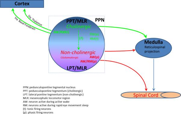
Forty-five years ago Shik and colleagues were the first to demonstrate that electrical stimulation of the dorsal pontine reticular formation induced fictive locomotion in decerebrate cats. This supraspinal motor site was subsequently termed the "mesencephalic locomotor region (MLR)". Cholinergic neurons of the pedunculopontine tegmental nucleus (PPT) have been suggested to form, or at least comprise in part, the neuroanatomical basis for the MLR, but direct evidence is lacking. In an effort to clarify the location and activity profiles of pontine reticulospinal neurons supporting locomotor behaviors, we employed in the present study a retrograde tracing method in combination with single-unit recordings and antidromic spinal cord stimulation as well as characterized the locomotor- and behavioral state-dependent activities of both reticulospinal and non-reticulospinal neurons. The retrograde labeling and antidromic stimulation responses suggested a candidate group of reticulospinal neurons that were non-cholinergic and located just medial to the PPT cholinergic neurons and ventral to the cuneiform nucleus (CnF). Unit recordings from these reticulospinal neurons in freely behaving animals revealed that the preponderance of neurons fired in relation to motor behaviors and that some of these neurons were also active during rapid eye movement sleep. By contrast, non-reticulospinal neurons, which likely included cholinergic neurons, did not exhibit firing activity in relation to motor behaviors. In summary, the present study provides neuroanatomical and electrophysiological evidence that non-cholinergic, pontine reticulospinal neurons may constitute the major component of the long-sought neuroanatomic MLR in mammals.
Copyright © 2012 IBRO. Published by Elsevier Ltd. All rights reserved.
Figures
 Filled blue (Phasic AW/REM);
  Open blue (Tonic AW/REM);
  Filled green (Phasic REM);
  Filled red (Phasic AW);
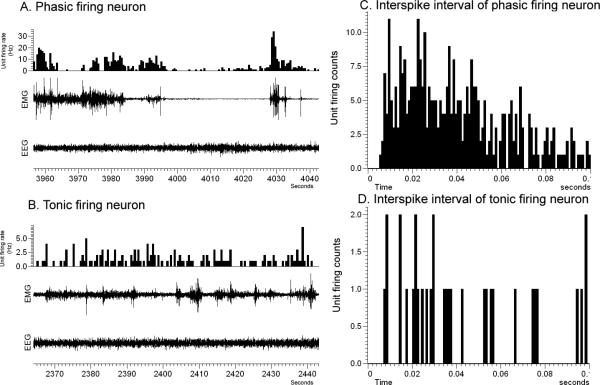
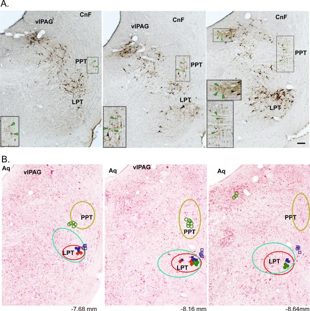
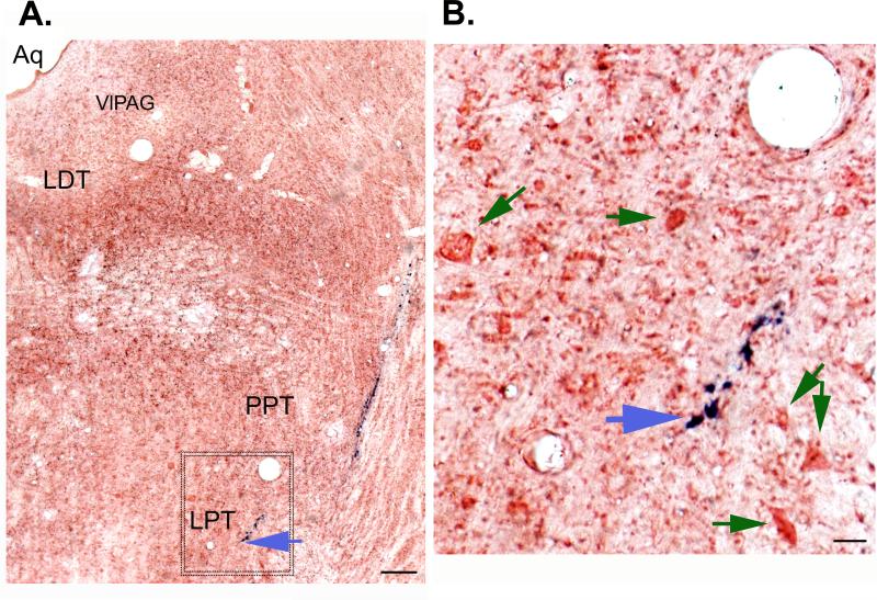
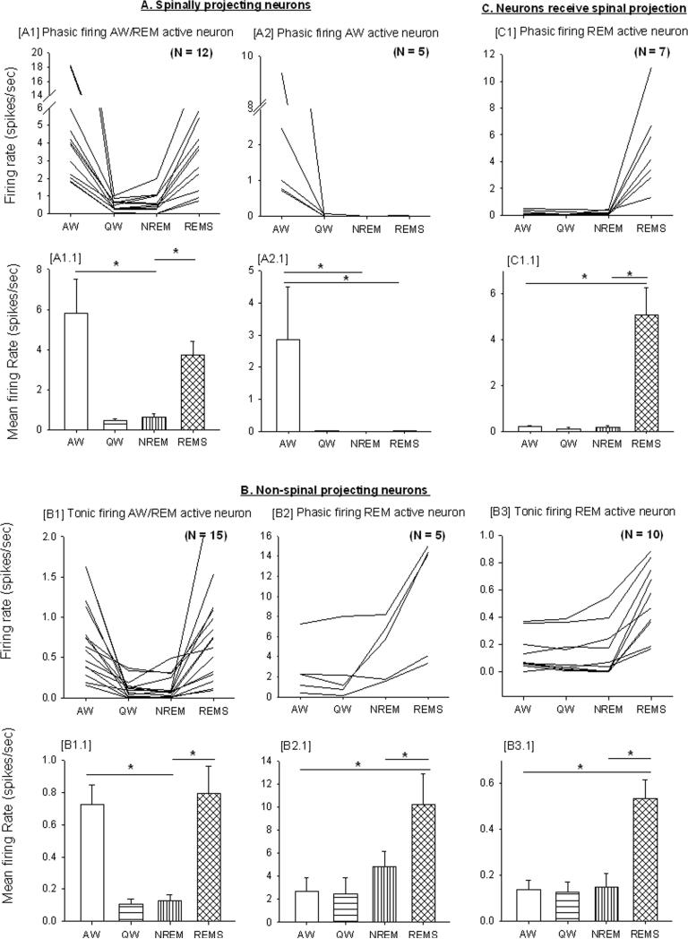
  Open green (Tonic REM); AW- Active wake active; AW/REM- Active wake and REM sleep active, REM-REM sleep active; Phasic – Phasic firing neurons show intermittent higher activity with occasional burst firing (usually preceded or followed by silent or low activity); Tonic – Tonic firing neurons show low or high firing activity but with regular interval. Numbers in B represent approximate AP distance (in mm) from bregma (Paxinos & Watson, 1998). The yellow circled region shows neurons with no relation to motor behavior nor antidromic response to spinal cord stimulation. Spinally-projecting neurons depicted here are CTb labeled cells (green circle) and neurons (red circle) with higher activity during motor behavior with antidromic projection to spinal cord. Bar in A. 50μm.
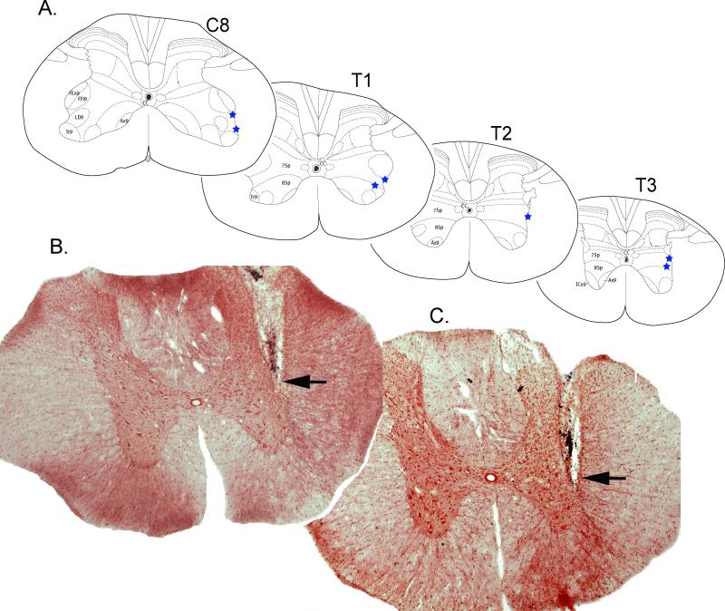
References
-
- Alam M, Schwabe K, Krauss JK. The pedunculopontine nucleus area: critical evaluation of interspecies differences relevant for its use as a target for deep brain stimulation. Brain. 2011;134:11–23. - PubMed
-
- Bernau NA, Puzdrowski RL, Leonard RB. Identification of the midbrain locomotor region and its relation to descending locomotor pathways in the Atlantic stingray, Dasyatis sabina. Brain Res. 1991;557:83–94. - PubMed
-
- Dormont JF, Conde H, Farin D. The role of the pedunculopontine tegmental nucleus in relation to conditioned motor performance in the cat. I. Context-dependent and reinforcement-related single unit activity. Exp Brain Res. 1998;121:401–410. - PubMed
Publication types
MeSH terms
Substances
Grants and funding
LinkOut - more resources
Full Text Sources
Miscellaneous
