. 2012 Sep 27:221:157-69.
doi: 10.1016/j.neuroscience.2012.07.006. Epub 2012 Jul 13.

Presynaptic facilitation of glutamate release in the basolateral amygdala: a mechanism for the anxiogenic and seizurogenic function of GluK1 receptors

Affiliations

Presynaptic facilitation of glutamate release in the basolateral amygdala: a mechanism for the anxiogenic and seizurogenic function of GluK1 receptors

V Aroniadou-Anderjaska et al. Neuroscience. .

Abstract

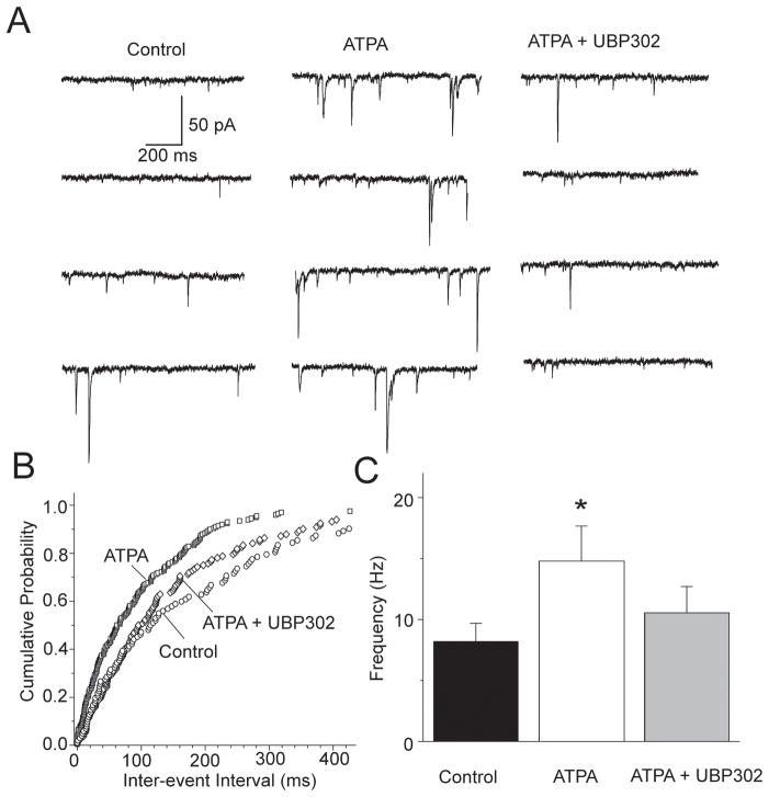
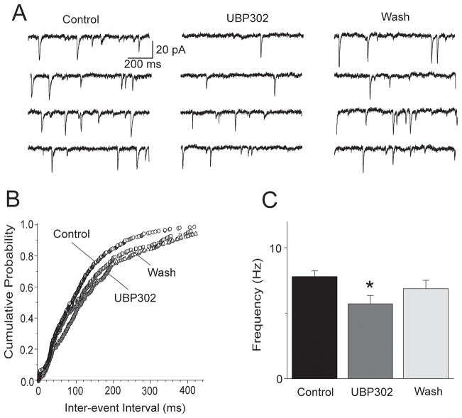
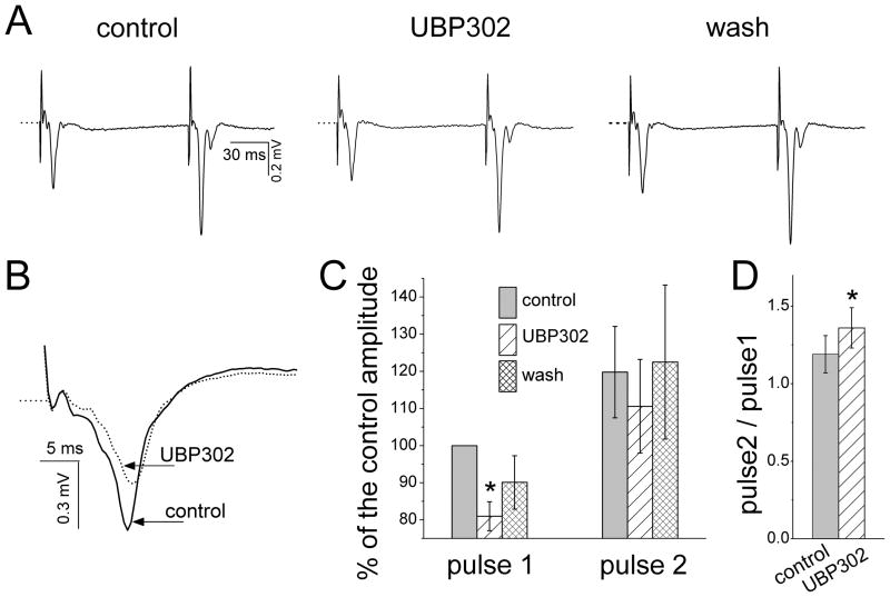
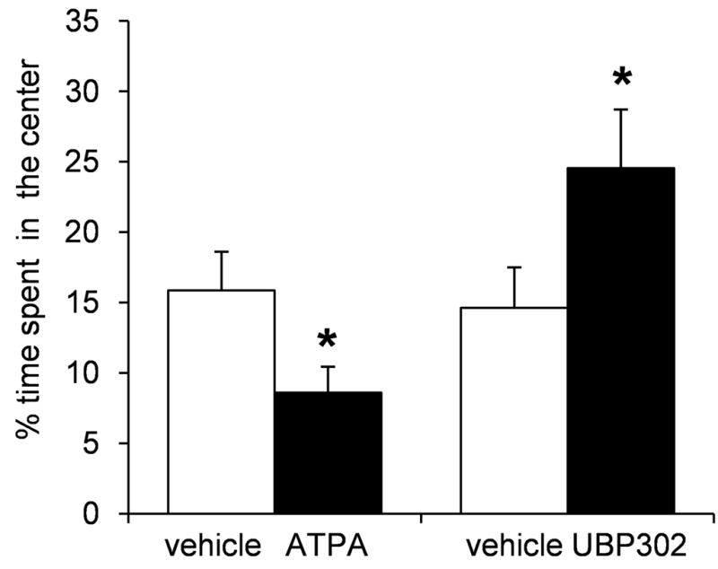
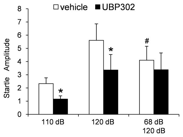
Kainate receptors containing the GluK1 subunit (GluK1Rs; previously known as GluR5 kainate receptors) are concentrated in certain brain regions, where they play a prominent role in the regulation of neuronal excitability, by modulating GABAergic and/or glutamatergic synaptic transmission. In the basolateral nucleus of the amygdala (BLA), which plays a central role in anxiety as well as in seizure generation, GluK1Rs modulate GABAergic inhibition via postsynaptic and presynaptic mechanisms. However, the role of these receptors in the regulation of glutamate release, and the net effect of their activation on the excitability of the BLA network are not well understood. Here, we show that in amygdala slices from 35- to 50-day-old rats, the GluK1 agonist (RS)-2-amino-3-(3-hydroxy-5-tert-butylisoxazol-4-yl) propanoic acid (ATPA) (300 nM) increased the frequency of spontaneous excitatory postsynaptic currents (sEPSCs) and miniature EPSCs (mEPSCs) recorded from BLA principal neurons, and decreased the rate of failures of evoked EPSCs. The GluK1 antagonist (S)-1-(2-amino-2-carboxyethyl)-3-(2-carboxybenzyl) pyrimidine-2,4-dione (UBP302) (25 or 30 μM) decreased the frequency of mEPSCs, reduced evoked field potentials, and increased the "paired-pulse ratio" of the field potential amplitudes. Taken together, these results suggest that GluK1Rs in the rat BLA are present on presynaptic terminals of principal neurons, where they mediate facilitation of glutamate release. In vivo bilateral microinjections of ATPA (250 pmol) into the rat BLA increased anxiety-like behavior in the open field test, while 2 nmol ATPA induced seizures. Similar intra-BLA injections of UBP302 (20 nmol) had anxiolytic effects in the open field and the acoustic startle response tests, without affecting pre-pulse inhibition. These results suggest that although GluK1Rs in the rat BLA facilitate both GABA and glutamate release, the facilitation of glutamate release prevails, and these receptors can have an anxiogenic and seizurogenic net function. Presynaptic facilitation of glutamate release may, in part, underlie the hyperexcitability-promoting effects of GluK1R activation in the rat BLA.


Figures

Fig. 1
Fig. 1
Activation of GluK1Rs by ATPA increases spontaneous EPSCs in principal BLA neurons. (A) Left: Rat brain slice showing the BLA (circle), where recordings were performed. Middle: The BLA nucleus at higher magnification; asterisk is marking the location of a patch-clamped neuron. Right: Patch-clamped pyramidal cell. Calibration bars: 1 mm, 500 μm, and 10 μm, from left to right, respectively. (B) Distinguishing BLA principal neurons from interneurons in current-clamp mode. Principal cells demonstrated variable patterns of accommodating spiking, while interneurons demonstrated fast, non-accomodating spiking. Responses were elicited by 500 ms-long current pulses, ranging from −300 pA to 300 pA with 100 pA steps. (C) Distinguishing BLA principal neurons from interneurons in voltage-clamp mode. Four 1s-long hyperpolarizing pulses starting from Vhold −70 mV to −80 mV and ending with −110 mV, elicited nonlinear current (Ih) in principal neurons and small linear current in interneurons. (D) Bath applied ATPA (300 nM) increased the frequency and amplitude of sEPSCs. Recordings are from a pyramidal-shaped neuron, in the presence of bicuculline (20 μM), D-AP5 (50 μM), and SCH50911 (20 μM). Example traces are shown in the left panel and group data from 7 cells in the bar graph to the right. *p < 0.05.
Fig. 2
Fig. 2
Activation of GluK1Rs by ATPA decreases the rate of failures of AMPA/kainate receptor-mediated synaptic currents recorded from principal neurons in the BLA. (A) Superimposed traces are eEPSCs recorded before, during, and after bath application of 300 nM ATPA. The slice medium contains D-AP5 (100 μM), bicuculline (20 μM), and SCH50911 (10 μM); Vhold, −70 mV. (B) Time course of the effect of ATPA on the amplitude of the eEPSCs and the number of failures (same cell as in A). (C) Pooled data of the percentage of failures (Mean ± SE) before, during, and after ATPA (n = 7; *p < 0.05).
Fig. 3
Fig. 3
The GluK1R agonist ATPA increases the frequency of mEPSCs. (A) Traces of mEPSCs recorded from a BLA pyramidal cell in control medium, during bath application of ATPA (300 nM), and after addition of UBP302 (30 μM). The slice medium contains bicuculline (20 μM), D-AP5 (100 μM), SCH50911 (20 μM), and TTX (1 μM); Vhold, −70 mV (B) Cumulative probability plot of inter-event intervals of mEPSCs in control medium, in the presence of ATPA, and in the presence of ATPA and UBP302 (data from the cell shown in A). (C) Group data showing the change in the frequency of mEPSCs by ATPA (n = 8, *p < 0.01).
Fig. 4
Fig. 4
The GluK1R antagonist UBP302 decreases the frequency of mEPSCs. (A) Traces of mEPSCs recorded from a BLA pyramidal neuron before, during, and after bath application of 30 μM UBP302. The slice medium contains bicuculline (20 μM), D-AP5 (100 μM), SCH50911 (20 μM), and TTX (1 μM); Vhold, −70 mV. (B) Cumulative probability plot of inter-event intervals of mEPSCs in control medium, in the presence of UBP302, and after washing out UBP302 (data from the cell shown in A). (C) Group data showing the change in the frequency of mEPSCs by UBP302 (n = 7, *p < 0.01).
Fig. 5
Fig. 5
Effects of UBP302 on field potentials evoked in the BLA by paired-pulse stimulation of the external capsule. (A) A representative example of BLA field synaptic responses to paired-pulse stimulation, before, during, and after wash out of bath-applied UBP302 (25 μM). Each trace is an average of 15 sweeps. Notice the reduction of the response to the first pulse by UBP302, without proportional reduction in the response to the second pulse. (B) An expanded view of field responses to the first stimulus pulse (different slice from the one shown in A), in control conditions and in the presence of UBP302 (red trace). Each of the two traces is the average of 15 sweeps. (C). Group data from 5 slices, showing the amplitude of the field responses to the first and the second stimulus pulses as percentages of the control response to the fist pulse, before, during, and after bath application of UBP302. Only the amplitude of the field response to the first stimulus pulse was significantly reduced by UBP302 (*p < 0.01). (D) Ratio of the amplitude of the synaptic response to the second pulse over the amplitude of the response to the first pulse, in control medium and in the presence of UBP302 (*p < 0.01).
Fig. 6
Fig. 6
Activation of GluK1Rs by bilateral intra-BLA injections of ATPA increases anxiety, while blockade of these receptors by UBP302 reduces anxiety, in the open field test. When rats were injected with ATPA (250 pmol), they spent significantly less time in the center of the open field compared to the time they spent in the center when injected with the vehicle. Conversely, when rats were injected with UBP302 (20 nmol), they spent significantly more time in the center of the open field compared to the time they spent in the center when injected with the vehicle. Bars show the Mean ± SE of the percentage of time spent in the center, from a group of 8 rats tested with ATPA or vehicle, and another group of 7 rats tested with UBP302 or vehicle. *p < 0.05
Fig. 7
Fig. 7
Blockade of GluK1Rs by bilateral intra-BLA injections of UBP302 reduces the amplitude of the acoustic startle response (ASR). The startle stimulus was 110 dB or 120 dB. In some trials, the 120 dB stimulus was preceded by a 68 dB prepulse (100 ms interval). The group data are Mean ± SE of the average ASR amplitude (arbitrary units) from 7 rats. UBP302 significantly reduced the amplitude of the ASR to both the 120 and 110 dB stimuli, but had no significant effect when the 120 dB noise burst stimuli were preceded by a prepulse. *p < 0.05, in comparison to the vehicle-injected rats; #p < 0.05 in comparison to the 120 dB stimulus that was not preceded by a prepulse.

References

    1. Alt A, Weiss B, Ogden AM, Li X, Gleason SD, Calligaro DO, Bleakman D, Witkin JM. In vitro and in vivo studies in rats with LY293558 suggest AMPA/kainate receptor blockade as a novel potential mechanism for the therapeutic treatment of anxiety disorders. Psychopharmacology (Berl) 2006;185:240–247. - PubMed
    1. Alt A, Weiss B, Ornstein PL, Gleason SD, Bleakman D, Stratford RE, Jr, Witkin JM. Anxiolytic-like effects through a GLUK5 kainate receptor mechanism. Neuropharmacology. 2007;52:1482–1487. - PubMed
    1. Apland JP, Aroniadou-Anderjaska V, Braga MF. Soman induces ictogenesis in the amygdala and interictal activity in the hippocampus that are blocked by a GluR5 kainate receptor antagonist in vitro. Neuroscience. 2009;159:380–389. - PMC - PubMed
    1. Aroniadou-Anderjaska V, Qashu F, Maria Braga MF. Mechanisms Regulating GABAergic Inhibitory Transmission in the Basolateral Amygdala: Implications for Epilepsy and Anxiety Disorders. Amino Acids. 2007;32:305–15. - PubMed
    1. Aroniadou-Anderjaska V, Fritsch B, Qashu F, Braga MF. Pathology and pathophysiology of the amygdala in epileptogenesis and epilepsy. Epilepsy Res. 2008;78:102–116. - PMC - PubMed

Publication types

MeSH terms