Review
. 2012 Aug;22(4):397-405.
doi: 10.1016/j.sbi.2012.06.002. Epub 2012 Jul 13.

Atomistic modeling of protein-DNA interaction specificity: progress and applications

Affiliations
Review

Atomistic modeling of protein-DNA interaction specificity: progress and applications

Limin Angela Liu et al. Curr Opin Struct Biol. 2012 Aug.

Abstract

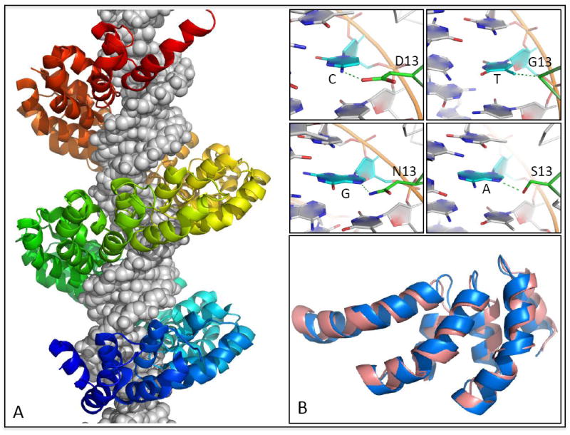
An accurate, predictive understanding of protein-DNA binding specificity is crucial for the successful design and engineering of novel protein-DNA binding complexes. In this review, we summarize recent studies that use atomistic representations of interfaces to predict protein-DNA binding specificity computationally. Although methods with limited structural flexibility have proven successful at recapitulating consensus binding sequences from wild-type complex structures, conformational flexibility is likely important for design and template-based modeling, where non-native conformations need to be sampled and accurately scored. A successful application of such computational modeling techniques in the construction of the TAL-DNA complex structure is discussed. With continued improvements in energy functions, solvation models, and conformational sampling, we are optimistic that reliable and large-scale protein-DNA binding prediction and engineering is a goal within reach.


Figures

Figure 1
Figure 1. The importance of flexibility in protein-DNA modeling
(A) Shifts in DNA bases (yellow arrows) are evident in template-based modeling of one zinc finger (the target, shown in red and pink) using another (the template, shown in blue). Interfaces are superimposed based on their protein components. Protein sidechains in the target whose DNA contacts would be disrupted by these DNA shifts are shown in stick representation (example taken from [7]). (B) Superposition of design model (pink) and experimental structure (blue) for the I-MsoI ‘-7C’ design of Ashworth et al. [47] reveals a shift of 2.9 Å in the DNA backbone and a 180° flip of a designed Tryptophan residue. These examples suggest that incorporation of backbone conformational flexibility may be necessary for reliable prediction and design of protein-DNA interactions.
Figure 2
Figure 2. Flowchart of structure-based calculation of the protein-DNA binding specificity
Step 1. A protein-DNA complex structure, taken from either experiments (X-ray or NMR) or molecular modeling (homology modeling or docking), serves as the input of the method. Here the Zif268 zinc finger protein is shown on the left in cartoon representation as an example (PDB ID: 1AAY). Step 2. The DNA base-pairs in the target site are mutated, perhaps exhaustively to all possible sequences (4L sequences for a site of length L), or more commonly to all single-base mutants (3L sequences). A superposition of four possible base pairs at a given position is shown in stick representation on the left. Step 3. The protein-DNA binding energy change caused by the DNA sequence mutation(s) is evaluated, using either a knowledge-based potential or a molecular mechanics force field. For methods that use molecular mechanics force fields, the binding energy difference is typically evaluated using the thermodynamic cycle shown on the left. Here the protein-DNA binding energy difference caused by the DNA sequence mutation (ΔΔG) is obtained by subtracting the unbound DNA energy (ΔGU) from the bound protein-DNA energy (ΔGB). Step 4. When single base pair mutations are performed, based on the approximation of DNA position independence, the binding energies of the different complexes can be converted into a position weight matrix and represented as a sequence logo [48] to show the binding specificity graphically (such as the logo shown on the left). When the DNA sequence space is exhaustively searched, a collection of high-affinity DNA sequences (such as those with a predicted binding affinity within some cutoff value of the optimal sequence) can also be represented as a sequence logo, although it should be noted that these sequences contain information regarding inter-position correlation that can be represented by higher-order graphical representations [49].
Figure 3
Figure 3. TAL effector-DNA interactions
(A) Structure of the 23.5-repeat TAL effector PthXo1 bound to its target site [42*] reveals the mechanistic basis of modular DNA recognition by TAL repeats. Successive repeats form a left-handed helical bundle that wraps around the DNA duplex paralleling the major groove. Inset panels show a subset of the specificity-determining contacts formed between repeat residue 13 and its associated base-pair in the target site. (B) A de novo TAL-effector model [*] is superimposed onto three repeats of the dHax3 [43*] TAL effector (1.2 Å Cα-RMSD over 102 residues). Whereas structural modeling played a critical role in structure determination for PthXo1, the dHax3 structure was solved by standard techniques and can serve as an independent reference for assessment of the structure predictions.

Similar articles

Cited by

References

    1. Zykovich A, Korf I, Segal DJ. Bind-n-Seq: high-throughput analysis of in vitro protein-DNA interactions using massively parallel sequencing. Nucleic Acids Research. 2009;37 - PMC - PubMed
    1. Robasky K, Bulyk ML. UniPROBE, update 2011: expanded content and search tools in the online database of protein-binding microarray data on protein-DNA interactions. Nucleic Acids Research. 2011;39:D124–D128. - PMC - PubMed
    1. Nutiu R, Friedman RC, Luo S, Khrebtukova I, Silva D, Li R, Zhang L, Schroth GP, Burge CB. Direct measurement of DNA affinity landscapes on a high-throughput sequencing instrument. Nature Biotechnology. 2011;29:659–U146. - PMC - PubMed
    1. Stormo GD, Zhao Y. Determining the specificity of protein-DNA interactions. Nature Reviews Genetics. 2010;11:751–760. A comprehensive review of state-of-the-art experimental techniques for determining protein-DNA binding specificity; both in vivo and in vitro methods are discussed. - PubMed
    1. Guerois R, Nielsen JE, Serrano L. Predicting changes in the stability of proteins and protein complexes: a study of more than 1000 mutations. J Mol Biol. 2002;320:369–387. - PubMed

Publication types

LinkOut - more resources