. 2012 Oct;33(29):7019-27.
doi: 10.1016/j.biomaterials.2012.06.052. Epub 2012 Jul 12.

The role of scaffold microarchitecture in engineering endothelial cell immunomodulation

Affiliations

The role of scaffold microarchitecture in engineering endothelial cell immunomodulation

Laura Indolfi et al. Biomaterials. 2012 Oct.

Abstract

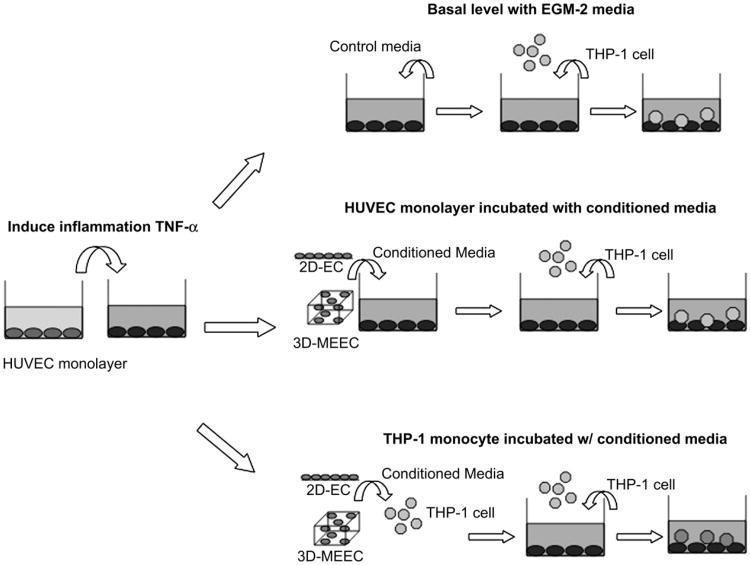
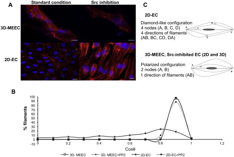
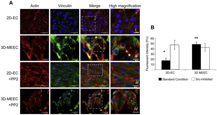
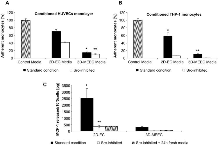
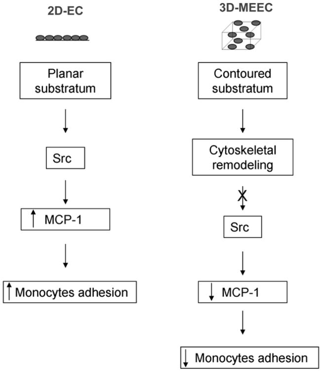
The implantation of matrix-embedded endothelial cells (MEECs) has been reported to have great therapeutic potential in controlling the vascular response to injury and maintaining patency in arteriovenous anastomoses. While there is an appreciation of their effectiveness in clinical and animal studies, the mechanisms through which they mediate these powerful effects remain relatively unknown. In this work, we examined the hypothesis that the 3-dimensional microarchitecture of the tissue engineering scaffold was a key regulator of endothelial behavior in MEEC constructs. Notably, we found that ECs in porous collagen scaffold had a markedly altered cytoskeletal structure with oriented actin fibers and rearrangement of the focal adhesion proteins in comparison to cells grown on 2D surfaces. We examined the immunomodulatory capabilities of MEECs and discovered that they were able to reduce the recruitment of monocytes to an inflamed endothelial monolayer by 5-fold compared to EC on 2D surfaces. An analysis of secreted factors from the cells revealed an 8-fold lower release of Monocyte Chemotactic Protein-1 (MCP-1) from MEECs. Differences between 3D and 2D cultured cells were abolished in the presence of inhibitors to the focal adhesion associated signaling molecule Src suggesting that adhesion-mediated signaling is essential in controlling the potent immunomodulatory effects of MEEC.


Conflict of interest statement

Authorship contributions and disclosure of conflicts of interest: L. Indolfi conceived the ideas, designed the research, performed experiments, analyzed data and wrote the paper. A.B. Baker and E.R. Edelman designed research, analyzed data and wrote the paper. The authors report no conflicts.

Figures

Fig. 1
Fig. 1
Experimental layout. A confluent endothelial monolayer (HUVECs) was activated by incubation with 10 ng/mL of TNF-α for 4 h. Top scheme: Thereafter, basal levels of THP-1 monocyte adhesion were analyzed by switching to EGM-2 media and incubate HUVEC monolayers with fluorescently labeled monocyte for 1 h before reading intensity of adherent cells. Two parallel sets of experiments were performed. Middle scheme: HUVECs were pre-incubated with conditioned media from flat 2D-ECs or contoured 3D-MEECs, then media changed to control EGM-2 and THP-1 added in suspension for 1 h before reading intensity of adherent cells. Bottom scheme: The same rational was pursued for monocytes, THP-1 cells were incubated with conditioned media from 2D-ECs or 3D-MEECs prior to use in the adhesion test.
Fig. 2
Fig. 2
Quantification of scaffold porosity. (A) Scheme of the layout used to determine scaffold pore size. 3D collagen-based scaffolds have been cryosectioned in 40 μm slide, stained with red dye and images recorded using a fluorescent microscope to determine the porosity of the mesh (i). Fluorescent micrographs were converted in binary images (ii), where each black area represented a pore (iii). Finally pore shape was approximated to an ellipsoid (iv) and the length of the major axis was evaluated. (B) Porosity analysis of the 3D collagen-based scaffold. Dimensions were grouped in range of 50 μm each ranging from 50 to 200 μm. The frequency of occurrence and the standard deviation of the analysis were then evaluated.
Fig. 3
Fig. 3
Microarchitecture of the substratum determines morphology and cytoskeleton rearrangement of ECs seeded in 2D and 3D domains. (A) Environmental-SEM micrographs of gelatin 3D scaffolds. (B) An individual struts at higher magnification. (C–D) 3D matrices seeded with ECs after 14 days induce a peculiar three-dimensional morphology of cells. (E–F) ECs seeded on planar 2D substrata attain their well-described cobblestone flat morphology. (G) Immunofluorescent images of ECs seeded on 3D matrixes better highlight the 3D contoured arrangement of cells induced by the underlying substratum: actin filaments (red), nuclei (blue) and gelatin matrix (green autofluorescence). (H) Cytoskeletal rearrangement is determined by the specific dimension of the cell substratum inducing the cell to wrap (insert i) or to bend (insert ii) around matrix struts. (I) The cytoskeleton of ECs on 2D is characterized by a diamond-like organization of the actin filaments. Scale bar: 3 mm (A); 100 μm (B); 50 μm (C,E,G,H,I); and 20 μm (D,F). (For interpretation of the references to color in this figure legend, the reader is referred to the web version of this article.)
Fig. 4
Fig. 4
Actin filaments orientation strongly depends on the substratum of culture and it is related to inhibition of Src pathway. (A) Fluorescent images of actin orientation for ECs cultured on 2D or 3D substrata both in standard condition and after inhibition of Src pathway (Scale bar is 10 μm for 3D-MEECs, and 20 μm for 2D-ECs). It is worth to notice that analysis of ECs within matrices was performed with the help of the 3D rendering of the z-stack data to reduce misjudgments due to planar projection of the different focal planes. (B) Distribution of actin filaments orientation in terms of angle θ spanned respect to a fixed direction, e.g. major axis in the cell, for 2D-ECs and 3D-MEEC in standard and Src-inhibited conditions (C) 2D-ECs exhibit under standard condition a peripheral dense distribution of the actin filaments. Cytoskeleton defines a diamond-like lattice, with four different directions of the fibers, mainly in the peripheral edges of the cell, and four connection points, e.g. nodes (top). Conversely 3D-MEECs are characterized by a more polarized configuration, with actin filaments predominantly parallel to the major axis of the cell. In this setting, fibers are oriented only in one direction with the presence of two nodes at the extremity (bottom). Inhibition of Src pathway determines such polarized configuration of the cytoskeleton in both settings (bottom). θ is the angle spanned between the filament and the major axis of the cells as depicted in the figure.
Fig. 5
Fig. 5
Substratum architecture of ECs culture induces changes in focal adhesion protein localization through inhibition of Src pathway. A) Immunofluorescent micrographs for ECs seeded in 2D or 3D settings: actin (red), vinculin (green) and nuclei (blue). Merged images show as vinculin is colocalized at the edge of actin fibers for flat 2D-ECs (i), while on contoured 3D-MEECs vinculin arrangement is perinuclear (ii). After incubation with a Src inhibitor (PP2) vinculin for 2D-ECs attains a perinuclear localization (iii) such as in the baseline 3D-MEECs (ii). Treatment of 3D-MEECs with PP2 does not significantly alter vinculin subcellular localization (iv). Scale bar: 50 μm. Inserts scale bar iiv: 25 μm. B) Vinculin quantification through an analysis of fluorescence intensity within single cell. Area of individual cell was selected and the intensity of the signal in the green channel was determined using the confocal software. 2D-ECs showed a mean signal of vinculin fluorescence that was 3-fold lower than the intensity from 3D-MEECs (*p < 0.05, versus all other groups). In contrast, when cells were incubated with Src inhibitor PP2, the signals among the two different cultures was identical and equal to the level of standard 3D-MEECs (**p > 0.05 versus Src-inhibited 2D and 3D settings). (For interpretation of the references to color in this figure legend, the reader is referred to the web version of this article.)
Fig. 6
Fig. 6
Substratum-induced ECs morphology affects biosecretory regulation of monocyte adhesion and depends upon Src inhibition. A) When flat 2D-ECs conditioned media (CM) was used to incubate vascular endothelial monolayer activated with TNF-α a reduction in monocytes adhesion of 30% respect to control was achieved. However, when contoured 3D-MEECs media was used, the percent inhibition of monocyte adherence increased to almost 5-fold. CM from flat 2D-ECs incubated with Src inhibitor PP2 reduced monocyte adhesion of over 50% respect to control. When Src-inhibited contoured 3D-MEECs media was used, a reduction higher than 80% was achieved (*p value < 0.005 versus all other groups; **p value > 0.05 versus standard 3D-MEEC). B) In a parallel set of experiments, in which CM was used to incubate monocytes prior to the adhesion test, a similar trend although with a higher percentage of adhesion inhibition with respect to control was shown. Both 2D- and 3D-MEECs treated with PP2 induced 90% less monocyte adhesion that control (*p value < 0.05 versus all other groups; **p value > 0.05 versus Src-inhibited 2D-EC). C) MCP-1 secretion by ECs is modulated by microarchitecture and requires Src signaling. ECs in 3D matrices release 8-fold less MCP-1 (289 ± 61 pg/105 cells) than in 2D culture (2526 ± 740 pg/105 cells). 24-h incubation with Src inhibitor PP2 drastically reduced secretion of MCP-1 either in flat 2D-ECs (328 ± 102 pg/105 cells) and contoured 3D-MEECs (30 ± 3 pg/105 cells). Incubation with fresh media for additional 24 h did not affect these levels (360 ± 28 versus 57 ± 9 pg/105 cells, 2D and 3D respectively). (*p value < 0.05 versus all other groups; **p value > 0.05 versus standard 3D-MEEC).
Fig. 7
Fig. 7
Scheme of the proposed pathway altered by the contoured subjacent surface sensed by the endothelial cells. The contoured topology of the substratum imposes a different cytoskeletal organization to ECs, which in turn interferes with the Src intracellular signaling. Reduction of secreted MCP-1 level, in turn, hinders monocytes adhesion to the site of inflammation. In contrast, in flat domain the Src signaling is not affected, therefore higher levels of MCP-1 and adherent monocytes are detected.

Similar articles

Cited by

References

    1. Ribatti D, Nico B, Vacca A, Roncali L, Dammacco F. Endothelial cell heterogeneity and organ specificity. J Hematother Stem Cell Res. 2002;11(1):81–90. - PubMed
    1. Michiels C. Endothelial cell functions. J Cell Physiol. 2003;196(3):430–43. - PubMed
    1. Nugent HM, Sjin RT, White D, Milton LG, Manson RJ, Lawson JH, et al. Adventitial endothelial implants reduce matrix metalloproteinase-2 expression and increase luminal diameter in porcine arteriovenous grafts. J Vasc Surg. 2007;46(3):548–56. - PMC - PubMed
    1. Nugent HM, Ng YS, White D, Groothius A, Kanner G, Edelman ER. Delivery site of perivascular endothelial cell matrices determines control of stenosis in a porcine femoral stent model. J Vasc Interv Radiol. 2009;20(12):1617–24. - PMC - PubMed
    1. Nugent HM, Rogers C, Edelman ER. Endothelial implants inhibit intimal hyperplasia after porcine angioplasty. Circ Res. 1999;84(4):384–91. - PubMed

Publication types