. 2012 Sep;181(3):993-1006.
doi: 10.1016/j.ajpath.2012.05.028. Epub 2012 Jul 13.

Interference with RhoA-ROCK signaling mechanism in autoreactive CD4+ T cells enhances the bioavailability of 1,25-dihydroxyvitamin D3 in experimental autoimmune encephalomyelitis

Affiliations

Interference with RhoA-ROCK signaling mechanism in autoreactive CD4+ T cells enhances the bioavailability of 1,25-dihydroxyvitamin D3 in experimental autoimmune encephalomyelitis

Ajaib S Paintlia et al. Am J Pathol. 2012 Sep.

Abstract

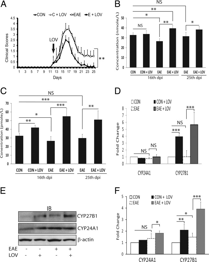
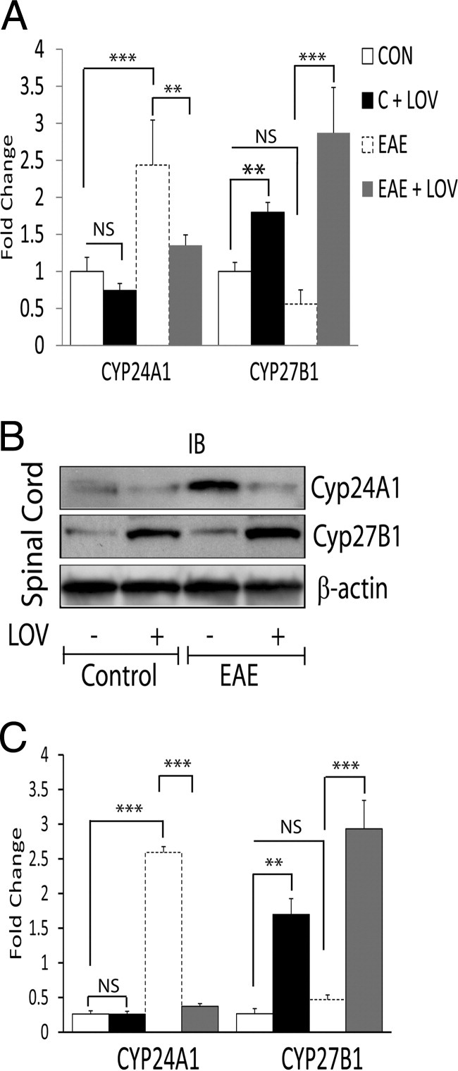
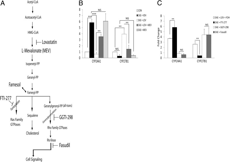
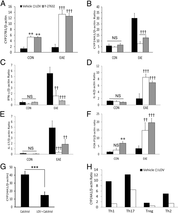
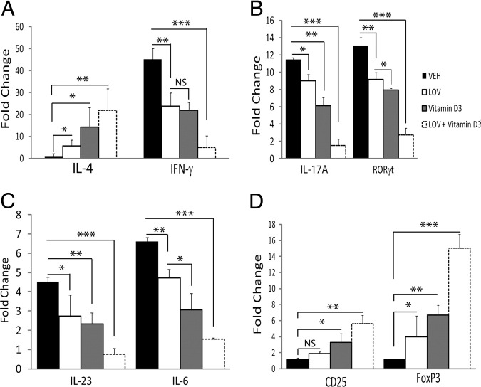
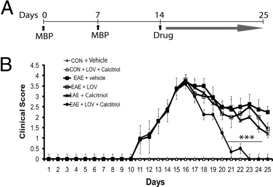
Vitamin D deficiency is a major risk factor for central nervous system (CNS) demyelinating diseases including multiple sclerosis (MS) and its animal model, that of experimental autoimmune encephalomyelitis (EAE). Both vitamin D(3) and 1, 25-dihydroxyviatmin-D(3) (calcitriol) had beneficial effects in EAE/MS. However, the exact cause of vitamin D deficiency in EAE/MS is not clear. Previously, we documented that lovastatin (LOV) provides protection in EAE animals via inhibition of RhoA-ROCK signaling. Herein, we demonstrate that LOV prevents the lowering of circulating 25-hydroxyvitamin-D(3) and 1,25-dihydroxyviatmin-D(3) levels including 1,25-dihydroxyviatmin-D(3) levels in the peripheral lymphoid organs and CNS of treated EAE animals. These effects of LOV were attributed to enhanced expression of vitamin D synthesizing enzyme (1α-hydroxylase) in kidney and the CNS, with corresponding reduction of vitamin D catabolizing enzyme (24-hydorxylase) expression in the CNS of EAE animals via inhibition of RhoA-ROCK signaling. Ex vivo and in vitro studies established that autoreactive Th1/Th17 cells had higher expression of 24-hydroxylase than Th2/T regulatory cells, that was reverted by LOV or ROCK inhibitor. Interestingly, LOV-mediated regulation of vitamin D metabolism had improved vitamin D(3) efficacy to confer protection in EAE animals and that was ascribed to the LOV- and calcitriol-induced immunomodulatory synergy. Together, these data provide evidence that interfering with RhoA-ROCK signaling in autoreactive Th1/Th17 cells can improve vitamin D(3) efficacy in clinical trials of MS and related neurodegenerative disorders.


Figures

Figure 1
Figure 1
Effect of LOV on clinical exacerbations, vitamin D metabolites, and expression of its metabolizing enzymes in EAE rats. EAE was induced in female Lewis rats with guinea pig MBP (30 μg/rat), and LOV (2 mg/kg) or vehicle treatments were initiated as described in Materials and Methods.A: Composite mean ± SEM of the clinical scores of 9 to 12 rats per group evaluated in three separate experiments. Composite mean ± SEM of four to five samples per group of the levels of 25-OH-D3 (B) and 1,25-(OH)2D3 (C) in the plasmas of treated EAE and control rats 16 dpi and 25 dpi. D: Composite mean ± SEM of four to five samples per group analyzed by real-time PCR in duplicate for CYP24A1 and CYP27B1 transcript levels in the kidneys of treated EAE and control rats 16 dpi. E: Representative autoradiograph of four samples per group depicting the levels of CYP27B1, CYP24A1, and β-actin proteins in the kidneys of treated EAE and control rats 16 dpi. F: Histogram depicts the composite mean ± SEM of four samples per group analyzed by Western blotting for CYP24A1 and CYP27B1 proteins normalized with β-actin and then compared with respective controls in the kidneys of treated EAE and control rats 16 dpi. Statistical significance as indicated: *P < 0.05; **P < 0.01; ***P < 0.001; NS, not significant.
Figure 2
Figure 2
Effect of LOV on CYP24A1 and CYP27B1 expression in the spinal cords of EAE rats. Spinal cords were collected 16 dpi from EAE and control rats treated with LOV or vehicle as detailed in Figure 1 legend. A: Composite mean ± SEM of four to five samples per group of the levels of CYP24A1 and CYP27B1 transcripts analyzed by real-time PCR in the spinal cord tissues. B: Representative autoradiograph depicting the levels of CYP24A1, CYP27B1, and β-actin proteins analyzed by Western blotting in the spinal cord tissues. C: Histogram depicting the composite mean ± SEM of four to five samples per group of the levels of CYP24A1 and CYP27B1 proteins normalized to β-actin and compared with respective controls in the spinal cord tissues. Statistical significance as indicated: **P < 0.01; ***P < 0.001; NS, not significant.
Figure 3
Figure 3
Effect of RhoA–ROCK signaling on the expressions of vitamin D–metabolizing enzymes in the spinal cords of EAE rats. EAE was induced in rats as detailed in the Figure 1 legend, and treatment with LOV alone or in combination with various metabolites of mevalonate pathway or specific enzyme inhibitor alone were initiated as described in Materials and Methods.A: Cartoon depicting the mevalonate pathway, and metabolites or enzyme inhibitors used to treat EAE rats are bolded. B: Plot depicts the levels of CYP24A1 and CYP27B1 transcripts analyzed by real-time PCR in duplicate in the spinal cords of EAE rats treated with LOV or metabolites of mevalonate pathway 16 dpi. C: Plot depicts the levels of CYP24A1 and CYP27B1 transcripts analyzed by real-time PCR in duplicate in the spinal cords of EAE and control rats treated with various inhibitors, ie, geranylgeranyl transferase (GGTI-298), farnesyl transferase (FTI-277) and RhoA kinase (fasudil). Results are expressed is the composite mean ± SEM from five rats per group of three separate experiments. Differences were statistical significance as indicated: **P < 0.01; ***P < 0.001; NS, not significant.
Figure 4
Figure 4
Effect of LOV and ROCK inhibitor on CYP24A1 and CYP27B1 expression in MBP-primed CD4+ T cells. Spleen cells were purified from the spleens of EAE rats by methods detailed under Materials and Methods. Spleen cells were stimulated with MBP (50 μg/mL) in the presence or absence of LOV (5 μmol/L). After 72 hours in culture, nonadhering cells were pelleted from the cultures and CD4+ T cells were purified using specific columns as described in Materials and Methods.A and B: Composite mean ± SEM for three to four samples analyzed in triplicate by real-time PCR for measuring the levels of CYP24A1 and CYP27B1 transcripts in MBP-sensitized CD4+ T cells. C–F: Composite mean ± SEM for IFN-γ, IL-4, IL-17, and FoxP3 transcripts levels in similarly treated MBP-sensitized CD4+ T cells. G: Composite mean ± SEM of three samples analyzed in triplicate by real-time PCR for the measurement of CYP24A1 transcripts in CD4+ T cells treated with calcitriol in the presence/absence of LOV for 24 hours. H: Composite mean of two samples per group analyzed in triplicate by real-time PCR to measure the levels of CYP24A1 transcripts in LOV-treated naive CD4+ T cells differentiated under Th1, Th17, Th2, and Treg polarizing conditions, as detailed in Materials and Methods. Differences were statistical significance as indicated: **P < 0.01 and not significant (NS) versus CON (vehicle); ††P < 0.01 and †††P < 0.001 versus EAE (vehicle); and ***P < 0.001 (G).
Figure 5
Figure 5
Effect of LOV and vitamin D3 therapy on EAE disease in rats fed a vitamin D3–deficient diet. Adult rats were fed a synthetic diet formulated to provide no vitamin D3 for 20 days before priming to MBP. EAE was induced and treatment with LOV (2 mg/kg, ip) and vitamin D3 (0.25 mg/kg, po) in combination or individually were started after the onset of disease. Composite mean ± SEM of three to four samples per group analyzed in triplicate by real-time PCR for IL-4 and IFN-γ (A), IL-17A and ROR-γt (B), IL-23 and IL-6 (C), and CD25 and FoxP3 (D) transcripts in the spinal cords of EAE and control rats treated with LOV or vitamin D3 16 dpi. Differences were statistical significance as indicated: *P < 0.05; **P < 0.01; ***P < 0.001. NS, not significant.
Figure 6
Figure 6
Effect of LOV and calcitriol combination on clinical exacerbation in EAE rats. EAE was induced in adult Lewis female rats as described in Figure 1 legend. Treatment with LOV (1 mg/kg) and calcitriol (1 μg/kg) was initiated individually or in combination in established EAE rats (14 dpi) and continued until the lessening of paralytic symptoms (25 dpi). A: Cartoon depicting the treatment strategy of EAE rats with LOV or calcitriol. B: Composite mean ± SEM of clinical scores of 10 to 12 rats per group evaluated in four separate experiments. LOV and calcitriol combination demonstrated significant lessening of paralytic symptoms 21 to 23 dpi and completely 25 dpi compared with their individual treatments or vehicle. Statistical significance as indicated: ***P < 0.001 versus EAE (vehicle).

Similar articles

Cited by

References

    1. Al-Omaishi J., Bashir R., Gendelman H.E. The cellular immunology of multiple sclerosis. J Leukoc Biol. 1999;65:444–452. - PubMed
    1. Prineas J.W., Barnard R.O., Kwon E.E., Sharer L.R., Cho E.S. Multiple sclerosis: remyelination of nascent lesions. Ann Neurol. 1993;33:137–151. - PubMed
    1. Bettelli E., Carrier Y., Gao W., Korn T., Strom T.B., Oukka M., Weiner H.L., Kuchroo V.K. Reciprocal developmental pathways for the generation of pathogenic effector TH17 and regulatory T cells. Nature. 2006;441:235–238. - PubMed
    1. Zhu J., Paul W.E. Peripheral CD4+ T-cell differentiation regulated by networks of cytokines and transcription factors. Immunol Rev. 2010;238:247–262. - PMC - PubMed
    1. Marrie R.A. Environmental risk factors in multiple sclerosis aetiology. Lancet Neurol. 2004;3:709–718. - PubMed

Publication types

MeSH terms