. 2012 Sep;143(3):708-718.e5.
doi: 10.1053/j.gastro.2012.05.053. Epub 2012 Jul 13.

Intestinal epithelial Toll-like receptor 4 regulates goblet cell development and is required for necrotizing enterocolitis in mice

Affiliations

Intestinal epithelial Toll-like receptor 4 regulates goblet cell development and is required for necrotizing enterocolitis in mice

Chhinder P Sodhi et al. Gastroenterology. 2012 Sep.

Abstract

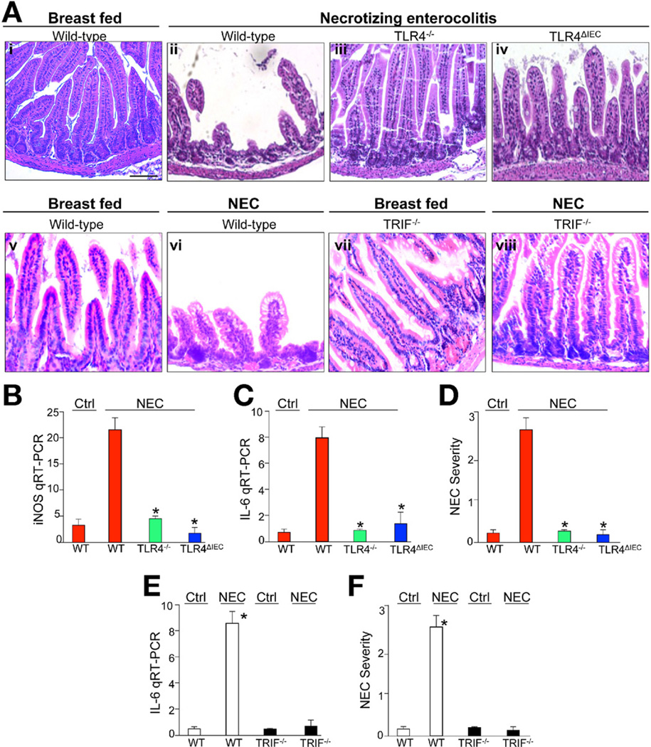
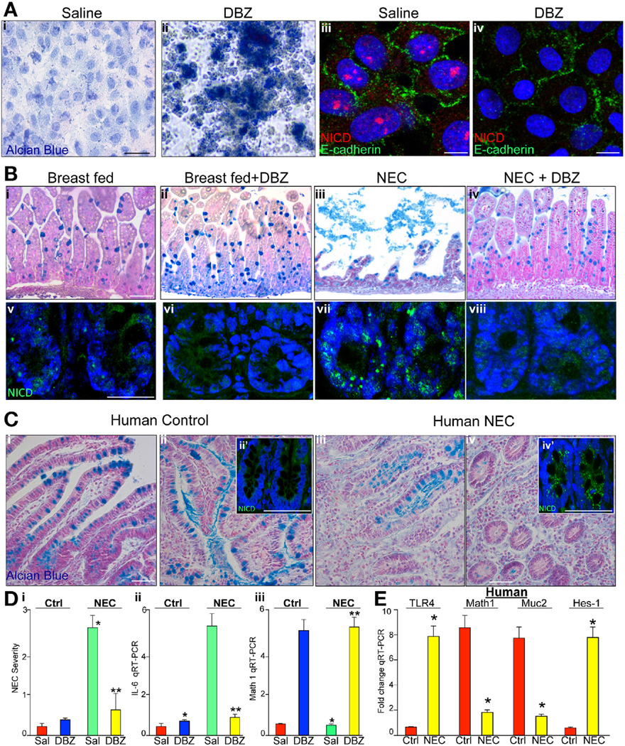
Background & aims: Little is known about factors that regulate intestinal epithelial differentiation; microbial recognition receptors such as Toll-like receptor (TLR)4 might be involved. We investigated whether intestinal TLR4 regulates epithelial differentiation and is involved in development of necrotizing enterocolitis (NEC) of the immature intestine.

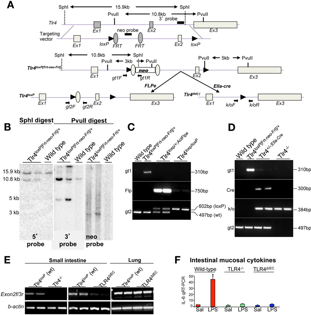
Methods: Mice with conditional disruption of TLR4 in the intestinal epithelium and TLR4 knockout (TLR4(-/-)) mice were generated by breeding TLR4(loxp/loxp) mice with villin-cre and Ella-cre, respectively. Enterocytes that did not express or overexpressed TLR4 were created by lentiviral or adenoviral transduction. Intestinal organoids were cultured on tissue matrices. Bile acids were measured by colorimetric assays, and microbial composition was determined by 16S pyrosequencing. NEC was induced in 7- to 10-day-old mice by induction of hypoxia twice daily for 4 days.

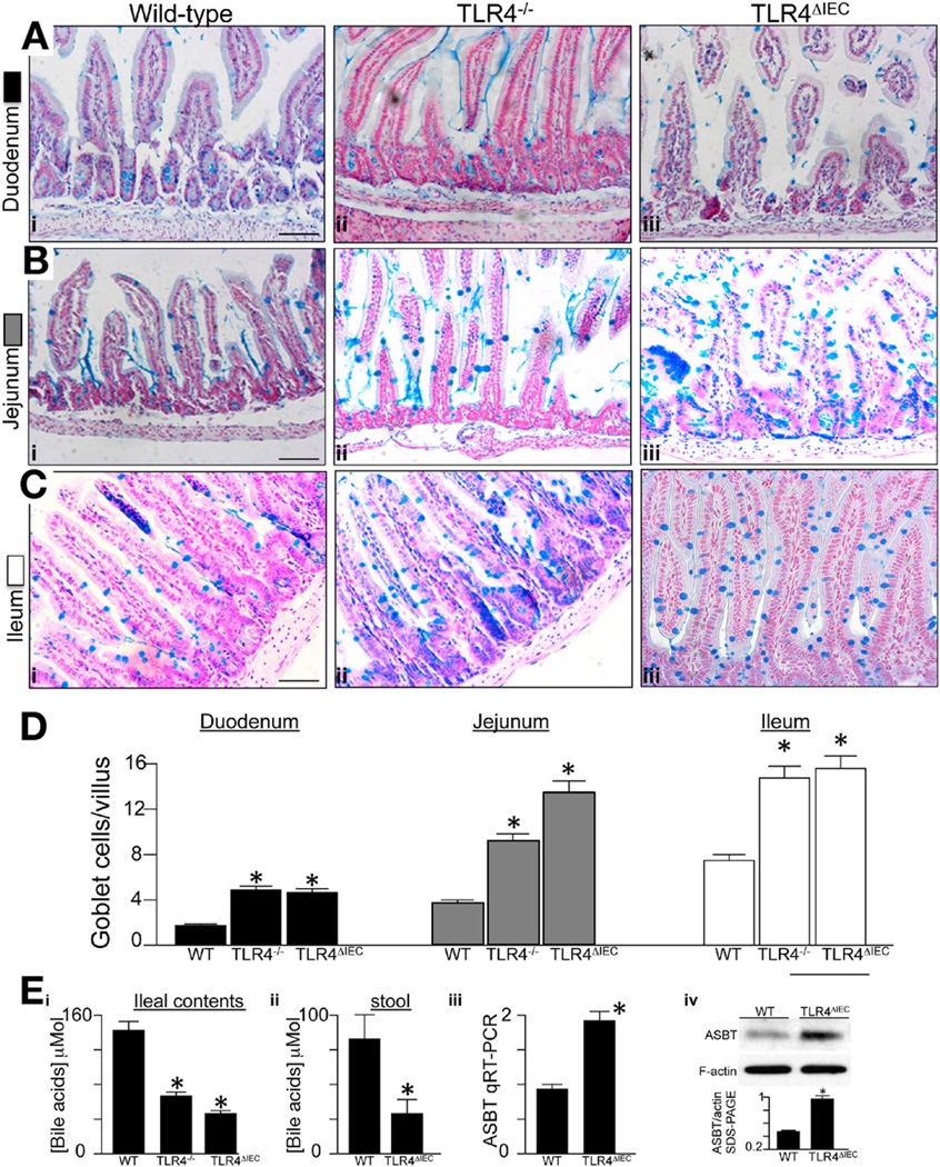
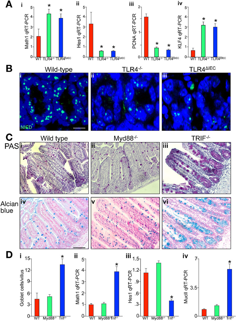
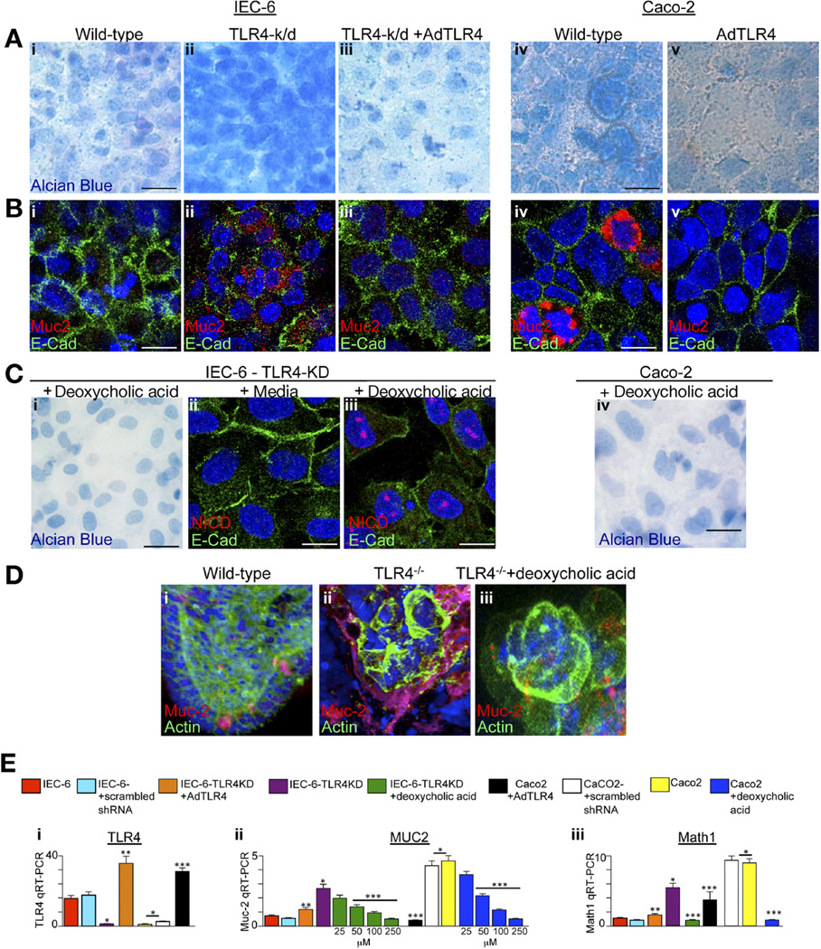
Results: TLR4(-/-) mice and mice with enterocyte-specific deletion of TLR4 were protected from NEC; epithelial differentiation into goblet cells was increased via suppressed Notch signaling in the small intestinal epithelium. TLR4 also regulates differentiation of goblet cells in intestinal organoid and enterocyte cell cultures; differentiation was increased on deletion of TLR4 and restored when TLR4 was expressed ectopically. TLR4 signaling via Notch was increased in intestinal tissue samples from patients with NEC, and numbers of goblet cells were reduced. 16S pyrosequencing revealed that wild-type and TLR4-deficient mice had similar microbial profiles; increased numbers of goblet cells were observed in mice given antibiotics. TLR4 deficiency reduced levels of luminal bile acids in vivo, and addition of bile acids to TLR4-deficient cell cultures prevented differentiation of goblet cells.

Conclusions: TLR4 signaling and Notch are increased in intestinal tissues of patients with NEC and required for induction of NEC in mice. TLR4 prevents goblet cell differentiation, independently of the microbiota. Bile acids might initiate goblet cell development.


Conflict of interest statement

Conflicts of interest

The authors disclose no conflicts.

Figures

Figure 1
Figure 1
Generation of TLR4loxp/loxp and TLR4-deficient mice. (A) Schematic diagram showing the generation of TLR4loxp/loxp mice. The positions of exons, neo-cassette, loxP, and Frt sites are shown along with restriction sites and positions of primers. (B) Southern blot showing the correct ligation of the targeting vector under conditions of the digest indicated. (C and D) Genomic PCR of tail-tip DNA confirming the genotype under the indicated conditions. (E) RT-PCR showing the correct excision of exon 2 messenger RNA in intestinal and lung mucosa under the indicated genetic conditions. (F) RT-PCR showing expression of IL-6 in the intestinal mucosa of wild-type (red bars), TLR4−/− mice (green bars), and TLR4ΔIEC mice (blue bars) 6 hours after injection of saline (Sal) or LPS (1 mg/kg intraperitoneally); *P < .05 versus saline, wild-type mice.
Figure 2
Figure 2
Deletion of TLR4 leads to increased goblet cells in the small intestine of mice. (AC) Alcian blue staining of (Aiiii) duodenum, (Biiii) jejunum, and (Diiii) ileum of (Ai, Bi, and Ci) wild-type C57/BL-6, (Aii, Bii, and Cii) TLR4−/−, and (Aii, Bii, and Cii) TLR4ΔIEC mice. Bars = 100 µm. (D) Quantification of goblet cells in the villi in more than 100 fields per group, averaged over at least 10 mice per group. (Ei and ii) Bile acid concentration, ileal contents, and stool in the indicated strain averaged over 10 mice/group; *P < .05 versus wild-type (WT); data are expressed as mean ± SEM. (Eiii and iv) Expression of ASBT by (iii) RT-PCR and (iv) sodium dodecyl sulfate/polyacrylamide gel electrophoresis (SDS-PAGE) in the indicated strain; *P < .05, 5 mice/group.
Figure 3
Figure 3
TLR4 regulates Notch signaling within the intestinal epithelium. (Aiiv) RT-PCR for (i) Math1, (ii) Hes1, (iii) PCNA, and (iv) KLF4 in wild-type (red bars), TLR4−/− (green bars), or TLR4ΔIEC (blue bars) mice. *P < .05 versus wild-type. Data are expressed as mean ± SEM. (Biiii) Confocal micrographs of terminal ileum stained for active Notch NICD (Notch intracellular domain). Bar = 10 µm. (Civi) Evaluation of goblet cells in the terminal ileum of (i and iv) wild-type mice, (ii and iv) Myd88−/− mice, and (iii and vi) TRIF−/− mice as stained by (iiii) periodic acid–Schiff and (ivvi) Alcian blue; quantification is shown in Di. Bar = 100 µm. Representative of 25 fields, at least 4 mice per field. (Diiiv) RT-PCR of (ii) Math1, (iii) Hes1, and (iv) MucII in wild-type (red), Myd88−/− (green), and TRIF−/− (blue) mice; *P < .05 versus wild-type. Data are expressed as mean ± SEM.
Figure 4
Figure 4
Deletion of TLR4 leads to increased goblet cell differentiation in cultured enterocytes. IEC-6 or Caco-2 cells that were wild-type (Ai and Bi, IEC-6; Aiv and Biv, Caco-2), deficient in TLR4 (Aii and Bii, TLR4-k/d), or deficient in TLR4 were infected with adenoviruses that express wild-type TLR4 (Aiii and Biii, TLR4-k/d + AdTLR4); Av and Bv show Caco-2 + AdTLR4, stained with (Aiv) Alcian blue or (Biv) Muc-2. E-cadherin is shown in green, DAPI in blue, and Muc-2 in red. (C) TLR4-deficient IEC-6 cells cultured for 5 days in (i and iii) deoxycholic acid or (ii) media and then stained for (i) Alcian blue or Notch intracellular signaling domain (red), E-cadherin (green), and DAPI (blue) in ii and iii. (Civ) Alcian blue staining in Caco-2 cells exposed to deoxycholic acid. (Diiii) Organoids from (i) wild-type, (ii) TLR4−/−, or (iii) TLR4−/− were then cultured in deoxycholic acid for (iii) 5 days and stained for DAPI (blue), actin (green), and Muc-2 (red). (Eiiii) RT-PCR in wild-type IEC-6 (red bars), IEC-6 cells transduced with scrambled shRNA (cyan bars), IEC-6–TLR4-KD (purple bars), IEC-6-TLR4-KD + AdTLR4 (orange bars), TLR4-KD + deoxycholic acid (green bars, at the indicated dose), wild-type Caco-2 cells (yellow bars), Caco-2 + AdTLR4 (black bars), Caco-2 + scrambled shRNA (white bars), and Caco-2 + deoxycholine (blue bars at the indicated dose). *P <.05 versus control IEC-6, **P <.05 versus IEC-6ΔIEC, ***P <.005 versus wild-type Caco-2. Data are expressed as mean ± SEM. Bar = 10 µm.
Figure 5
Figure 5
TLR4 in the intestinal epithelium is required for the development of NEC. Terminal ileum from newborn mice and evaluated by H&E after (Ai, v, and vii) breastfeeding or (Aiiiv, vi, viii) induction of NEC in either TLR4 or TRIF-deficient strains. (B, C, and E) RT-PCR in intestinal mucosa of wild-type (red bars or white bars), TLR4−/− (green bars), TLR4ΔIEC (blue bars), and TRIF−/− (black bars) for (B) inducible nitric oxide synthase or (C and E) IL-6 as shown. (D and F) Severity of NEC. Mean ± SEM; **P < .05 versus wild-type (WT) NEC; *P <.05 versus WT control. Representative of >100 fields, >10 mice/group. Bar = 100 µm.
Figure 6
Figure 6
Inhibition of Notch leads to increased goblet cells and reduced severity of NEC. (Ai and ii) IEC-6 cells in (i) absence or (ii) presence of DBZ stained by Alcian blue. (Aiii and iv) Confocal micrographs of IEC-6 cells stained for E-cadherin (green) or active Notch1, that is, NICD (red in iii and iv). (Biviii) Alcian blue (iiv) or confocal for active Notch (NICD, green) and DAPI (blue) in terminal ileum of newborn mice (vviii). Bar = 100 µm; representative of more than 10 mice/group in 3 separate experiments. (Ciiv) Terminal ileum of human infants undergoing surgical resection of the intestine for (iii and iv) NEC or (i and ii) controls; ii? and iv?: NICD. Representative of more than 10 patients/group. (Di) Severity of NEC. (Dii and iii) RT-PCR for (ii) IL-6 and (iii) Math1. For Diiii: *P < .05 versus saline breastfed; **P < .05 versus saline NEC. *P < .05, yellow versus red. Bar = 100 µm. Data are expressed as mean ± SEM. (E) RT-PCR for TLR4, Math1, Muc-2, and hes1 as indicated in human ileum from control patients (red) and patients with NEC (yellow); *P < .05 versus control; 15 patients per group.

References

    1. van der Flier LG, Clevers H. Stem cells, self-renewal, and differentiation in the intestinal epithelium. Annu Rev Physiol. 2009;71:241–260. - PubMed
    1. Neal MD, Leaphart C, Levy R, et al. Enterocyte TLR4 mediates phagocytosis and translocation of bacteria across the intestinal barrier. J Immunol. 2006;176:3070–3079. - PubMed
    1. Cario E, Rosenberg IM, Brandwein SL, et al. Lipopolysaccharide activates distinct signaling pathways in intestinal epithelial cell lines expressing Toll-like receptors. J Immunol. 2000;164:966–972. - PubMed
    1. Neu J, Walker WA. Necrotizing enterocolitis. N Engl J Med. 2011;363:255–264. - PMC - PubMed
    1. Jilling T, Simon D, Lu J, et al. The roles of bacteria and TLR4 in rat and murine models of necrotizing enterocolitis. J Immunol. 2006;177:3273–3282. - PMC - PubMed

Publication types

MeSH terms