

Aging-associated shifts in functional status of mast cells located by adult and aged mesenteric lymphatic vessels
- PMID: 22796537
- PMCID: PMC3468454
- DOI: 10.1152/ajpheart.00378.2012
Aging-associated shifts in functional status of mast cells located by adult and aged mesenteric lymphatic vessels
Abstract
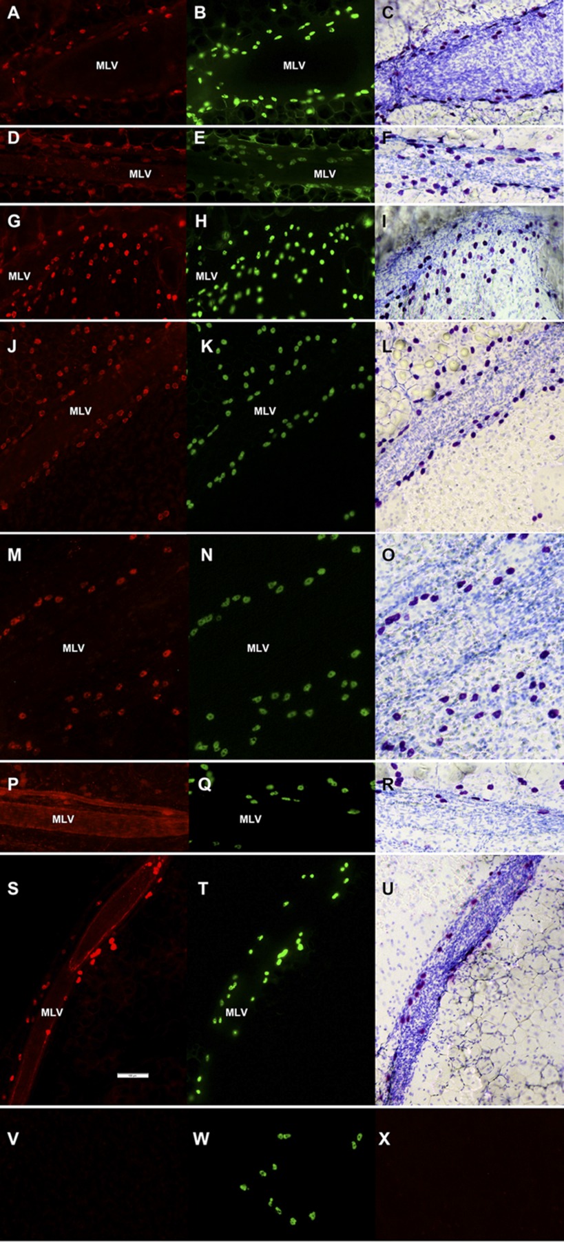
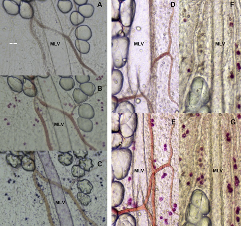
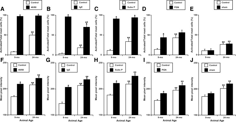
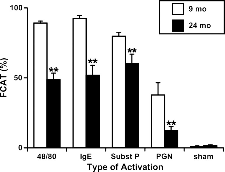
We had previously proposed the presence of permanent stimulatory influences in the tissue microenvironment surrounding the aged mesenteric lymphatic vessels (MLV), which influence aged lymphatic function. In this study, we performed immunohistochemical labeling of proteins known to be present in mast cells (mast cell tryptase, c-kit, prostaglandin D(2) synthase, histidine decarboxylase, histamine, transmembrane protein 16A, and TNF-α) with double verification of mast cells in the same segment of rat mesentery containing MLV by labeling with Alexa Fluor 488-conjugated avidin followed by toluidine blue staining. Additionally, we evaluated the aging-associated changes in the number of mast cells located by MLV and in their functional status by inducing mast cell activation by various activators (substance P; anti-rat DNP Immunoglobulin E; peptidoglycan from Staphyloccus aureus and compound 48/80) in the presence of ruthenium red followed by subsequent staining by toluidine blue. We found that there was a 27% aging-associated increase in the total number of mast cells, with an ∼400% increase in the number of activated mast cells in aged mesenteric tissue in resting conditions with diminished ability of mast cells to be newly activated in the presence of inflammatory or chemical stimuli. We conclude that higher degree of preactivation of mast cells in aged mesenteric tissue is important for development of aging-associated impairment of function of mesenteric lymphatic vessels. The limited number of intact aged mast cells located close to the mesenteric lymphatic compartments to react to the presence of acute stimuli may be considered contributory to the aging-associated deteriorations in immune response.
Figures
References
-
- Amin K. The role of mast cells in allergic inflammation. Respir Med 106: 9–14, 2012 - PubMed
-
- Beil WJ, Schulz M, Wefelmeyer U. Mast cell granule composition and tissue location—a close correlation. Histol Histopathol 15: 937–946, 2000 - PubMed
-
- Benoit JN, Zawieja DC. Gastrointestinal lymphatics. In: Physiology of the gastrointestinal tract, edited by Johnson L. New York: Raven Press, 1994, p. 1669–1692
-
- Bergstresser PR, Tigelaar RE, Tharp MD. Conjugated avidin identifies cutaneous rodent and human mast cells. J Invest Dermatol 83: 214–218, 1984 - PubMed
Publication types
MeSH terms
Substances
Grants and funding
LinkOut - more resources
Full Text Sources
Medical
Research Materials