

Mapping cardiac surface mechanics with structured light imaging
- PMID: 22796539
- PMCID: PMC3468459
- DOI: 10.1152/ajpheart.00269.2012
Mapping cardiac surface mechanics with structured light imaging
Abstract
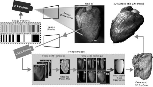
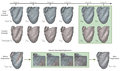
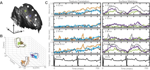
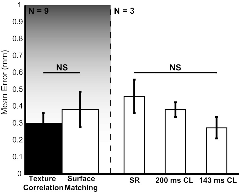
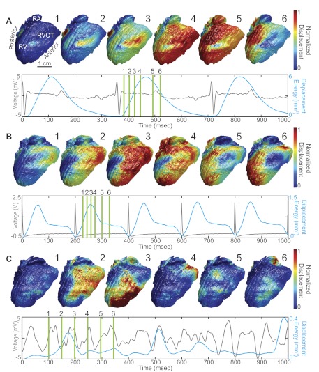
Cardiovascular disease often manifests as a combination of pathological electrical and structural heart remodeling. The relationship between mechanics and electrophysiology is crucial to our understanding of mechanisms of cardiac arrhythmias and the treatment of cardiac disease. While several technologies exist for describing whole heart electrophysiology, studies of cardiac mechanics are often limited to rhythmic patterns or small sections of tissue. Here, we present a comprehensive system based on ultrafast three-dimensional (3-D) structured light imaging to map surface dynamics of whole heart cardiac motion. Additionally, we introduce a novel nonrigid motion-tracking algorithm based on an isometry-maximizing optimization framework that forms correspondences between consecutive 3-D frames without the use of any fiducial markers. By combining our 3-D imaging system with nonrigid surface registration, we are able to measure cardiac surface mechanics at unprecedented spatial and temporal resolution. In conclusion, we demonstrate accurate cardiac deformation at over 200,000 surface points of a rabbit heart recorded at 200 frames/s and validate our results on highly contrasting heart motions during normal sinus rhythm, ventricular pacing, and ventricular fibrillation.
Figures
Similar articles
-
Electromechanical wavebreak in a model of the human left ventricle.Am J Physiol Heart Circ Physiol. 2010 Jul;299(1):H134-43. doi: 10.1152/ajpheart.00862.2009. Epub 2010 Apr 16. Am J Physiol Heart Circ Physiol. 2010. PMID: 20400690
-
Three-dimensional visualization of phase singularities on the isolated rabbit heart.J Cardiovasc Electrophysiol. 2002 Dec;13(12):1311. doi: 10.1046/j.1540-8167.2002.01311.x. J Cardiovasc Electrophysiol. 2002. PMID: 12521354 No abstract available.
-
The importance of non-uniformities in mechano-electric coupling for ventricular arrhythmias.J Interv Card Electrophysiol. 2014 Jan;39(1):25-35. doi: 10.1007/s10840-013-9852-0. Epub 2013 Dec 12. J Interv Card Electrophysiol. 2014. PMID: 24338157 Review.
-
Organization of ventricular fibrillation in the human heart: experiments and models.Exp Physiol. 2009 May;94(5):553-62. doi: 10.1113/expphysiol.2008.044065. Epub 2009 Jan 23. Exp Physiol. 2009. PMID: 19168541
-
Transmural dispersion of repolarization and arrhythmogenicity: the Brugada syndrome versus the long QT syndrome.J Electrocardiol. 1999;32 Suppl:158-65. doi: 10.1016/s0022-0736(99)90074-2. J Electrocardiol. 1999. PMID: 10688320 Review.
Cited by
-
Putting the pieces together using in vivo optical mapping.Cardiovasc Res. 2019 Sep 1;115(11):1574-1575. doi: 10.1093/cvr/cvz089. Cardiovasc Res. 2019. PMID: 30924872 Free PMC article. No abstract available.
-
Toward panoramic in situ mapping of action potential propagation in transgenic hearts to investigate initiation and therapeutic control of arrhythmias.Front Physiol. 2014 Sep 8;5:337. doi: 10.3389/fphys.2014.00337. eCollection 2014. Front Physiol. 2014. PMID: 25249982 Free PMC article.
-
Optical Mapping of Membrane Potential and Epicardial Deformation in Beating Hearts.Biophys J. 2016 Jul 26;111(2):438-451. doi: 10.1016/j.bpj.2016.03.043. Biophys J. 2016. PMID: 27463145 Free PMC article.
-
3D absolute shape measurement of live rabbit hearts with a superfast two-frequency phase-shifting technique.Opt Express. 2013 Mar 11;21(5):5822-32. doi: 10.1364/OE.21.005822. Opt Express. 2013. PMID: 23482151 Free PMC article.
-
Optical Mapping of Cardiac Electromechanics.Biophys J. 2016 Jul 26;111(2):269-270. doi: 10.1016/j.bpj.2016.04.052. Biophys J. 2016. PMID: 27463128 Free PMC article. No abstract available.
References
-
- Allen B, Curless B. The space of human body shapes: reconstruction and parameterization from range scans. ACM T Graphic 22: 587–594, 2003
-
- Bogun F, Taj M, Ting M, Kim HM, Reich S, Good E, Jongnarangsin K, Chugh A, Pelosi F, Oral H, Morady F. Spatial resolution of pace mapping of idiopathic ventricular tachycardia/ectopy originating in the right ventricular outflow tract. Heart Rhythm 5: 339–344, 2008 - PubMed
-
- Bray MA, Lin SF, Wikswo JP. Three-dimensional surface reconstruction and fluorescent visualization of cardiac activation. IEEE Trans Biomed Eng 47: 1382–1391, 2000 - PubMed
Publication types
MeSH terms
Grants and funding
LinkOut - more resources
Full Text Sources
Medical