. 2012 Sep 1;303(5):H523-32.
doi: 10.1152/ajpheart.00826.2011. Epub 2012 Jul 13.

Role of matrix Gla protein in angiotensin II-induced exacerbation of vascular calcification

Affiliations

Role of matrix Gla protein in angiotensin II-induced exacerbation of vascular calcification

Guanghong Jia et al. Am J Physiol Heart Circ Physiol. .

Erratum in

  • Corrigendum.
    [No authors listed] [No authors listed] Am J Physiol Heart Circ Physiol. 2016 Apr 1;310(7):H951. doi: 10.1152/ajpheart.zh4-1873-corr.2016. Am J Physiol Heart Circ Physiol. 2016. PMID: 27036402 Free PMC article. No abstract available.

Abstract

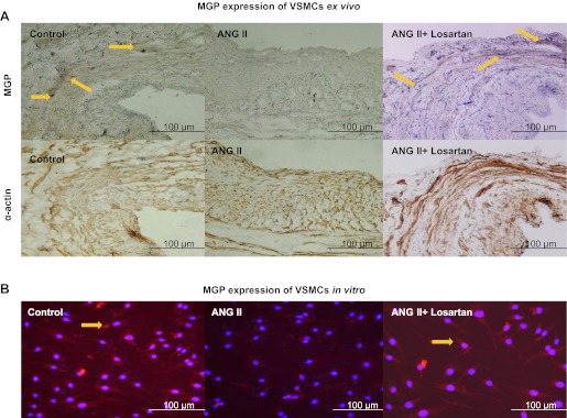
Vascular calcification predicts an increased risk for cardiovascular events in atherosclerosis, diabetes, and end-stage kidney diseases. Matrix Gla protein (MGP), an inhibitor of calcification, limits calcium phosphate deposition in the vessel wall. There are many factors contributing to the progression of atherosclerosis, including hypertension, hyperlipidemia, the renin-angiotensin system, and inflammation. Angiotensin II (ANG II) plays a crucial role in the atherogenic process through not only its pressor responses but also its growth-promoting and inflammatory effects. In this study, we investigated the role of MGP in ANG II-induced exacerbation of vascular calcification in human vascular smooth muscle cells (VSMCs). The expression of MGP, calcification, and apoptosis in human VSMCs were examined by Western blot analysis, real-time PCR, in situ terminal deoxynucleotidyltransferase-mediated dUTP nick end labeling, and enzyme-linked immunosorbent assay, respectively. Increase in VSMC calcification in human atherosclerotic plaques upregulates MGP expression and apoptosis in a negative feedback manner. ANG II inhibited MGP expression in VSMCs via and in vitro in a dose- and time-dependent manner through ANG II type 1 receptor and NF-κB signaling pathway. Meanwhile, MGP inhibited the calcification, caspase-3 activity, activation of runt-related transcription factor 2, and release of inflammatory cytokines by VSMCs induced by calcification medium (2.5 mM P(i)) and ANG II in vitro. These observations provide evidence that ANG II exacerbates vascular calcification through activation of the transcription factors, runt-related transcription factor 2 and NF-κB, and regulation of MGP, inflammatory cytokines expression in human VSMCs.


Figures

Fig. 1.
Fig. 1.
The association between calcification, matrix Gla protein (MGP) expression, and apoptosis on vascular smooth muscle cells (VSMCs) from human carotid plaque and normal carotid artery. The calcification, MGP expression, and apoptosis were checked by von Kossa staining, immunohistochemistry, and in situ terminal deoxynucleotidyltransferase-mediated dUTP nick end labeling, respectively. Histological serial adjacent sections were taken from human carotid plaque and normal carotid artery. Arrows indicate the calcification, MGP expression, and apoptosis positive cells, which were clearly visible in smooth muscle α-actin positive cells.
Fig. 2.
Fig. 2.
ANG II-induced inhibition of MGP expression in VSMCs via and in vitro. A: histological serial adjacent sections were taken from the ANG II (10−7 mol/l)-treated human saphenous vein with or without losartan (10−6 mol/l) for 24 h. Arrows indicate MGP expression positive cells, which were clearly visible in smooth muscle α-actin positive cells. B: VSMCs were stimulated by ANG II (10−7 mol/l) with or without losartan (10−6 mol/l) for 24 h, and expression of MGP was determined by immunostaining. Arrows indicate MGP expression (red) in cultured SMCs, and the nuclear was stained with blue color in 4′,6′-diamidino-2-phenylindole.
Fig. 3.
Fig. 3.
ANG II-induced inhibition of MGP mRNA expression through ANG II type-1 receptor (AT1R) in human VSMCs. A: ANG II (10−7 mol/l) inhibited MGP mRNA expression of VSMCs in a time-dependent manner. B: ANG II inhibited MGP mRNA expression of VSMCs at 24 h in a dose-dependent manner. C: IC50 of ANG II in the downregulation of MGP mRNA was 1.29 × 10⁻⁷ mol/l. D: ANG II receptor antagonist losartan (10−6 mol/l, AT1R receptor specific) and PD-123319 (10−6 mol/l, ANG II type-2 receptor specific) were added to the cultured medium for 30 min and exposed to 10−7 mol/l ANG II for an additional 24 h. E: calcification medium (2.5 mM Pi) induced MGP mRNA expression of VSMCs in vitro. Each bar represents the 3 independent experiments. #P < 0.01 compared with control group; *P < 0.01 compared with ANG II group.
Fig. 4.
Fig. 4.
Involvement of the activation of transcription factor NF-κB in ANG II-inhibited MGP expression in human VSMCs. A: ANG II (10−7 mol/l) induced NF-κB p65 nuclear translocation at 5, 15, and 30 min in VSMCs. Arrows indicate that NF-κB p65 were clearly visible in the part of VSMC nuclear. B: ANG II (10−7 mol/l) induced nuclear protein expression of NF-κB in VSMCs. C: VSMCs were stimulated by ANG II with or without NF-κB inhibitor II (20 nmol/l), and mRNA expression of MGP was determined by real-time PCR. Data are means ± SE of 3 independent experiments. #P < 0.01 compared with control group; *P < 0.01 compared with ANG II group.
Fig. 5.
Fig. 5.
Effect of MGP on the calcification and caspase-3 activity induced by phosphate and ANG II in VSMCs. A: MGP inhibited the calcification of VSMCs induced by phosphate and ANG II. The VSMC calcification was checked by Alizarin Red-S. Arrows indicate the calcification of positive cells. B: overexpression of MGP in VSMCs using pN-FLAG-hMGP vector at 24 and 48 h. C: the calcium content of smooth muscle cells was measured by o-cresolphthalein complexone, normalized by cellular protein content. D: MGP inhibited the caspase-3 activity of VSMC induced by phosphate and ANG II. OD405, optical density at 405 nm. E: ANG II induced the expression of bone morphogenic protein-2 (BMP-2) in VSMCs. Data are means ± SE of 3 independent experiments. #P < 0.01 compared with control group; *P < 0.05 compared with 2.5 mM Pi group; †P < 0.01 compared with 2.5 mM Pi group; ‡P < 0.01 compared with 2.5 mM Pi + ANG II group.
Fig. 6.
Fig. 6.
The effect of MGP and NF-κB inhibitor II on the activation of runt-related transcription factor 2 (Runx2) and expression of IL-6, IL-8, and monocyte chemoattractant protein-1 (MCP-1) in human VSMCs. A: MGP inhibited ANG II-induced the activation of transcription factor Runx2 in human VSMCs. B: MGP inhibited phosphate-medium plus ANG II-induced activation of transcription factor Runx2 in human VSMCs. C: MGP inhibited ANG II-induced protein expression of IL-6, IL-8, and MCP-1 in VSMCs. D: MGP and NF-κB inhibitor II (20 nmol/l) inhibited phosphate-medium plus ANG II-induced protein expression of IL-6, IL-8, and MCP-1 in VSMCs. #P < 0.01 compared with control group; †P < 0.01 compared with 2.5 mM Pi group; *P < 0.01 compared with ANG II group or 2.5 mM Pi + ANG II group.
Fig. 7.
Fig. 7.
Mechanisms depicted ANG II-induced exacerbation of vascular calcification. ANG II exacerbates the vascular calcification through activation of the transcription factor Runx2 and NF-κB and regulation of MGP, inflammatory cytokines, and BMP-2 expression in human VSMCs.

Similar articles

Cited by

References

    1. Burdon KP, Rudock ME, Lehtinen AB, Langefeld CD, Bowden DW, Register TC, Liu Y, Freedman BI, Carr JJ, Hedrick CC, Rich SS. Human lipoxygenase pathway gene variation and association with markers of subclinical atherosclerosis in the diabetes heart study. Mediators Inflamm 2010: 170153, 2010 - PMC - PubMed
    1. Hu WY, Fukuda N, Satoh C, Jian T, Kubo A, Nakayama M, Kishioka H, Kanmatsuse K. Phenotypic modulation by fibronectin enhances the angiotensin II-generating system in cultured vascular smooth muscle cells. Arterioscler Thromb Vasc Biol 20: 1500–1505, 2000 - PubMed
    1. Jia G, Aggarwal A, Yohannes A, Gangahar DM, Agrawal DK. Cross-talk between angiotensin II and IGF-1-induced connexin 43 expression in human saphenous vein smooth muscle cells. J Cell Mol Med 15: 1695–1702, 2011 - PMC - PubMed
    1. Jia G, Cheng G, Gangahar DM, Agrawal DK. Involvement of connexin 43 in angiotensin II-induced migration and proliferation of saphenous vein smooth muscle cells via the MAPK-AP-1 signaling pathway. J Mol Cell Cardiol 44: 882–890, 2008 - PMC - PubMed
    1. Johnson RC, Leopold JA, Loscalzo J. Vascular calcification: pathobiological mechanisms and clinical implications. Circ Res 99: 1044–1059, 2006 - PubMed

Publication types

MeSH terms