. 2012 Sep;53(3):437-45.
doi: 10.1016/j.yjmcc.2012.07.001. Epub 2012 Jul 13.

Cardiac specific ATP-sensitive K+ channel (KATP) overexpression results in embryonic lethality

Affiliations

Cardiac specific ATP-sensitive K+ channel (KATP) overexpression results in embryonic lethality

Amir Toib et al. J Mol Cell Cardiol. 2012 Sep.

Abstract

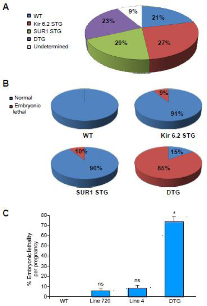
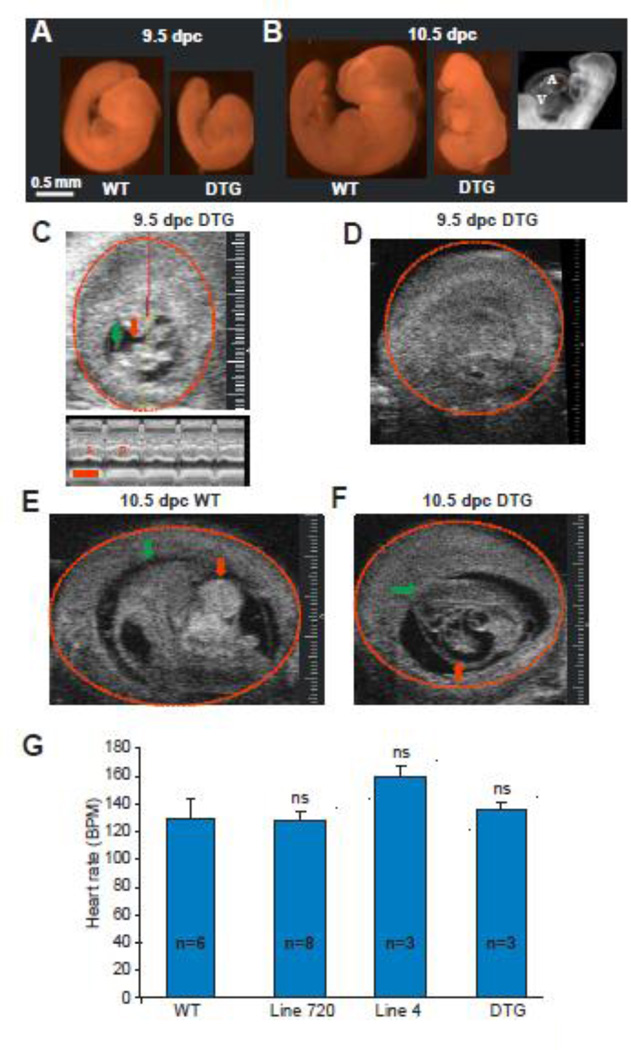
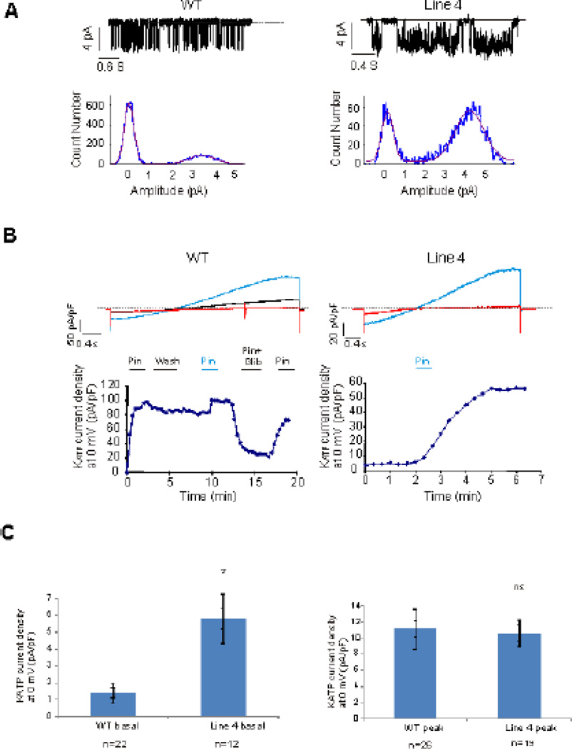
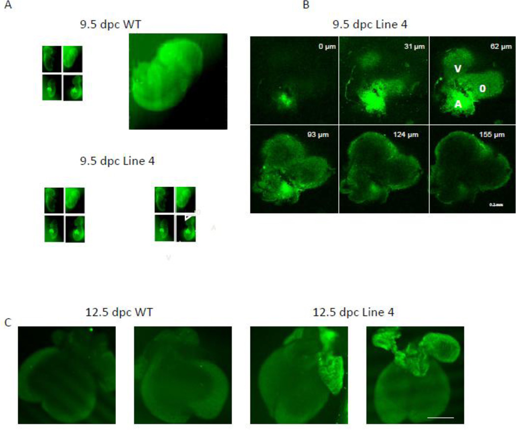
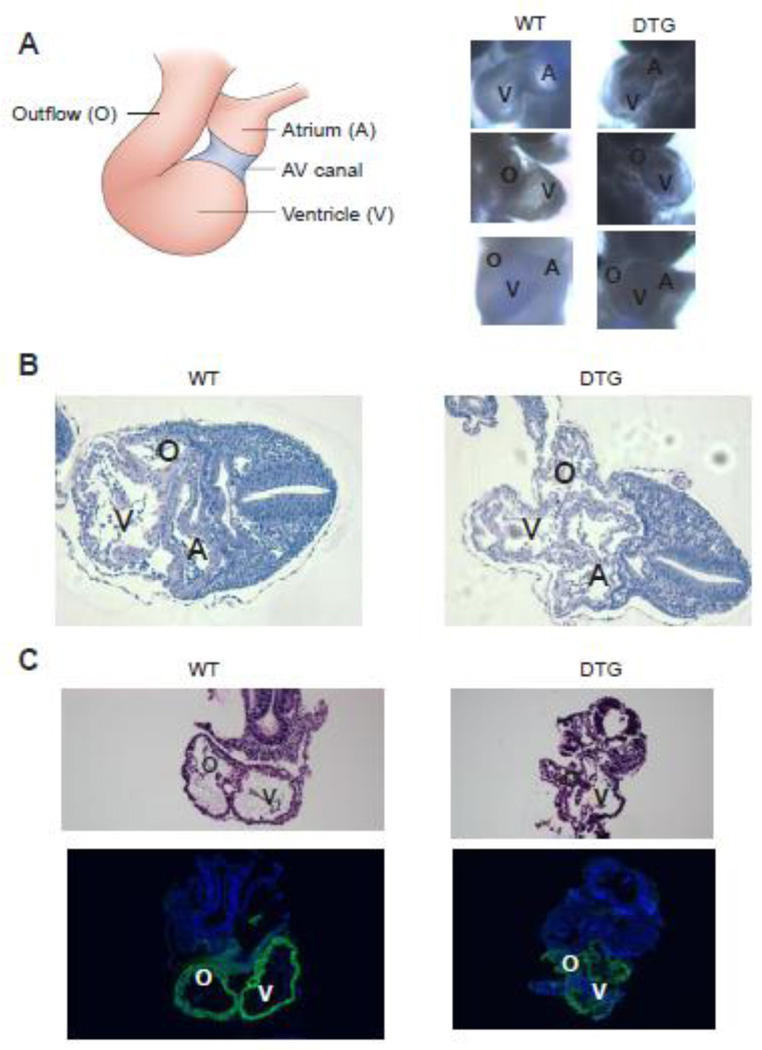
Transgenic mice overexpressing SUR1 and gain of function Kir6.2[∆N30, K185Q] K(ATP) channel subunits, under cardiac α-myosin heavy chain (αMHC) promoter control, demonstrate arrhythmia susceptibility and premature death. Pregnant mice, crossed to carry double transgenic progeny, which harbor high levels of both overexpressed subunits, exhibit the most extreme phenotype and do not deliver any double transgenic pups. To explore the fetal lethality and embryonic phenotype that result from K(ATP) overexpression, wild type (WT) and K(ATP) overexpressing embryonic cardiomyocytes were isolated, cultured and voltage-clamped using whole cell and excised patch clamp techniques. Whole mount embryonic imaging, Hematoxylin and Eosin (H&E) and α smooth muscle actin (αSMA) immunostaining were used to assess anatomy, histology and cardiac development in K(ATP) overexpressing and WT embryos. Double transgenic embryos developed in utero heart failure and 100% embryonic lethality by 11.5 days post conception (dpc). K(ATP) currents were detectable in both WT and K(ATP)-overexpressing embryonic cardiomyocytes, starting at early stages of cardiac development (9.5 dpc). In contrast to adult cardiomyocytes, WT and K(ATP)-overexpressing embryonic cardiomyocytes exhibit basal and spontaneous K(ATP) current, implying that these channels may be open and active under physiological conditions. At 9.5 dpc, live double transgenic embryos demonstrated normal looping pattern, although all cardiac structures were collapsed, probably representing failed, non-contractile chambers. In conclusion, K(ATP) channels are present and active in embryonic myocytes, and overexpression causes in utero heart failure and results in embryonic lethality. These results suggest that the K(ATP) channel may have an important physiological role during early cardiac development.


Figures

Figure 1
Figure 1. KATP overexpression results in embryonic lethality
(A) Genotype distribution from breeding of heterozygous STG mice overexpressing either SUR1 line 720 or gain of function Kir6.2(ΔN30, K185Q) line 4. 20 pregnancies were assessed between 9.5–18 dpc (with a total of 166 embryos). Embryonic genotyping distributed as expected from a heterozygous inheritance pattern of each of the single transgenes, with ~ 1/4 of the embryos of each genotype. (B) Embryonic lethality distribution of the different genotypes. Of note, the 15% fraction for DTG (right lower pie), represents embryos that were alive (beating heart) at 9.5–10.5 dpc, none of the DTG was alive at 11.5 dpc. (C) Embryonic lethality per pregnancy from the above crosses (* p<0.05 compared to WT, Line 720 and Line 4). No embryonic lethality was detected in 20 WT embryos. All of the DTG embryos extracted after 11.5 dpc were dead.
Figure 2
Figure 2. KATP overexpression causes in utero heart failure and embryonic lethality
(A) Littermate 9.5 dpc WT (left) and DTG (right) embryos. DTG embryo demonstrates growth retardation. (B) 10.5 dpc WT (left) and DTG (middle) embryos. DTG embryo demonstrates growth retardation, pericardial effusion and diffuse hydrops. Dark field imaging (right) demonstrates that the 10.5 dpc DTG heart is arrested developmentally at the primitive loop stage (9.5dpc). (C) Two dimensional ultrasound image of a 9.5 dpc live DTG embryo with a normally beating heart. The head is marked with a green arrow. The heart is marked with a red arrow. The embryonic sac is marked with a red circle. The red/yellow line represents the M mode plane across the embryo’s heart. The lower M mode panel demonstrates systole (S) and diastole (D). Sizing scale (for C–E) indicates 0.1 mm. Timing bar (red) in lower panel indicates 200 msec. (D) Two dimensional ultrasound of a 9.5 dpc DTG embryo, a littermate of the embryo in C, demonstrating an empty sac with an absorbed embryo. (E) Two dimensional ultrasound image of a 10.5 dpc WT embryo with normal development. (F) Two dimensional ultrasound of a 10.5 dpc DTG embryo demonstrating growth retarded embryo with pericardial effusion. (G) Summary of fetal heart rates (BPM=beats per minute) in live embryos demonstrates no statistical difference between the different genotype groups.
Figure 3
Figure 3. KATP currents are detected in embryonic myocytes throughout development
(A) Single channel KATP currents recorded in excised patches from 15 dpc WT (left) and 12 dpc Line 4 (right) ventricular myocytes. Current density histograms demonstrate 3.5–4 pA single channel current, corresponding to a 70–80 pS single channel conductance. (B) Whole cell voltage-clamp recordings of 12 dpc WT and 18 dpc Line 4 ventricular myocytes (Upper panels). Red: baseline current. Blue: Maximal KATP activation in presence of 100µM of Pinacidil. Black: current in presence of 10µM Glibenclamide and 100µM Pinacidil. (Lower panels) KATP current versus time (Pin=Pinacidil, Glib=Glibenclamide). (C) Whole cell basal (left) and peak (right) KATP current density in WT and Line 4 embryonic cardiomyocytes. Basal KATP current was significantly increased in Line 4 cardiomyocytes compared to early cardiac development WT cardiomyocytes (p<0.05) No difference was noted in peak KATP current between WT and line 4 cardiomyocytes.
Figure 4
Figure 4. Gain of function KATP transgene is mainly expressed in atrial tissue
(A) Confocal images of two 9.5 dpc WT (control) littermate embryos (upper two images) and two 9.5 Line 4 littermate embryos hearts (lower 2 images). Note prominent GFP fluorescence in Line 4 transgenic mice throughout the hearts. A=atrium, V=ventricle, O=outflow. Sizing bar indicates 1 mm. (B) Z-sectioning (with depth information in the right upper corner of each image) confocal images of the heart from 9.5 dpc Line 4 embryo (right lower embryo in A). A=atrium, V=ventricle, O=outflow. Sizing bar indicates 0.2 mm. (C) Confocal images of two 12.5 dpc WT (control) littermate embryonic hearts (left 2 images) and two 12.5 Line 4 littermate embryonic hearts (right 2 images). Sizing bar indicates 1 mm.
Figure 5
Figure 5. DTG embryos demonstrate normal looping pattern
(A) Left: Schematic frontal view of 9.5 dpc looped heart. Right: Bright field images of 9.5 dpc WT and DTG embryos demonstrate normal looping pattern. Upper panel: left lateral view. Middle panel: right lateral view. Lower panel: frontal view. A=atrium, V=ventricle, O=outflow. (B) H&E staining of transverse sections of 9.5 dpc WT (left) and DTG embryos. Note that although looping pattern was normal, DTG embryos demonstrate collapse of all cardiac structures. A=atrium, V=ventricle, O=outflow. (C) H&E staining of sagittal sections of 9.5 dpc WT (left) and DTG. Lower panel demonstrates positive immunostaining (green) for α-smooth muscle actin (α-SMA), a marker of early cardiac mesoderm, in both WT and DTG. V=ventricle, O=outflow.

Similar articles

Cited by

References

    1. Zhang H, Flagg TP, Nichols CG. Cardiac sarcolemmal K(ATP) channels: Latest twists in a questing tale! J Mol Cell Cardiol. Jan;48(1):71–75. - PMC - PubMed
    1. Kane GC, Liu XK, Yamada S, Olson TM, Terzic A. Cardiac KATP channels in health and disease. J Mol Cell Cardiol. 2005 Jun;38(6):937–943. - PMC - PubMed
    1. Zingman LV, Alekseev AE, Hodgson-Zingman DM, Terzic A. ATP-sensitive potassium channels: metabolic sensing and cardioprotection. J Appl Physiol. 2007 Nov;103(5):1888–1893. - PubMed
    1. Olson TM, Terzic A. Human K(ATP) channelopathies: diseases of metabolic homeostasis. Pflugers Arch. Jul;460(2):295–306. - PMC - PubMed
    1. Tong X, Porter LM, Liu G, Dhar-Chowdhury P, Srivastava S, Pountney DJ, et al. Consequences of cardiac myocyte-specific ablation of KATP channels in transgenic mice expressing dominant negative Kir6 subunits. Am J Physiol Heart Circ Physiol. 2006 Aug;291(2):H543–H551. - PMC - PubMed

Publication types