. 2012 Sep 15;53(6):1213-21.
doi: 10.1016/j.freeradbiomed.2012.07.003. Epub 2012 Jul 14.

Activation of AMP-activated protein kinase alleviates high-glucose-induced dysfunction of brain microvascular endothelial cell tight-junction dynamics

Affiliations

Activation of AMP-activated protein kinase alleviates high-glucose-induced dysfunction of brain microvascular endothelial cell tight-junction dynamics

Chao Liu et al. Free Radic Biol Med. .

Abstract

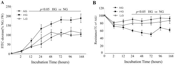
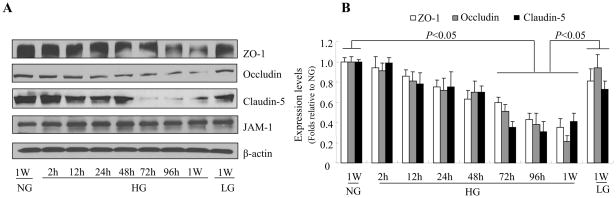
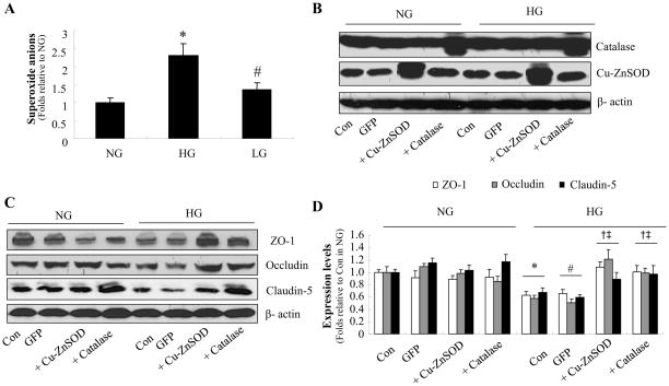
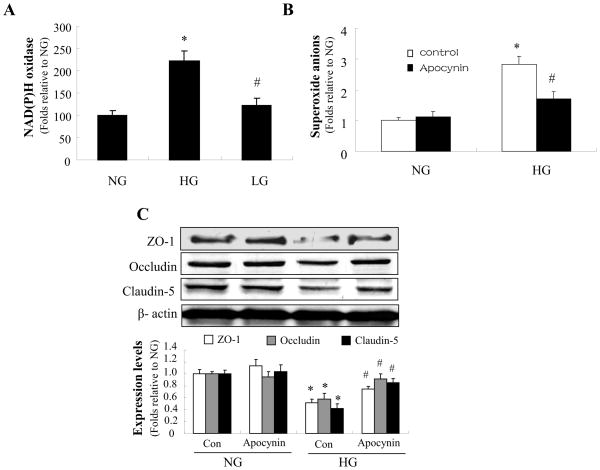
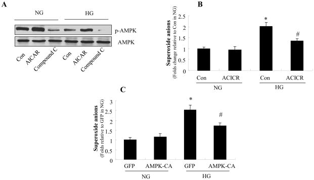
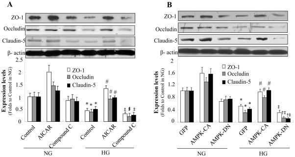
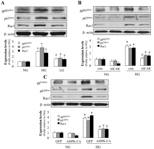
The blood-brain barrier, formed by specialized brain endothelial cells that are interconnected by tight junctions, strictly regulates paracellular permeability to maintain an optimal extracellular environment for brain homeostasis. Diabetes is known to compromise the blood-brain barrier, although the underlying mechanism remains unknown. The aim of this study was to elucidate the molecular mechanisms underlying disruption of the blood-brain barrier in diabetes and to determine whether activation of AMP-activated protein kinase prevents diabetes-induced blood-brain barrier dysfunction. Exposure of human brain microvascular endothelial cells to high glucose (25 mmol/L D-glucose), but not to high osmotic conditions (20 mmol/L L-glucose plus 5 mmol/L D-glucose), for 2h to 1 week significantly increased the permeability of the blood-brain barrier in parallel with lowered expression levels of zonula occludens-1, occludin, and claudin-5, three proteins that are essential to maintaining endothelial cell tight junctions. In addition, high glucose significantly increased the generation of superoxide anions. Adenoviral overexpression of superoxide dismutase or catalase significantly attenuated the high-glucose-induced reduction of endothelial cell tight-junction proteins. Furthermore, administration of apocynin reversed the effects of high glucose on endothelial cell tight-junction proteins. Finally, activation of AMP-activated protein kinase with 5-amino-4-imidazole carboxamide riboside or adenoviral overexpression of constitutively active AMP-activated protein kinase mutants abolished both the induction of NAD(P)H oxidase-derived superoxide anions and the tight-junction protein degradation induced by high glucose. We conclude that high glucose increases blood-brain barrier dysfunction in diabetes through induction of superoxide anions and that the activation of AMP-activated protein kinase protects the integrity of the blood-brain barrier by suppressing the induction of NAD(P)H oxidase-derived superoxide anions.


Figures

Figure 1
Figure 1. High glucose decreases blood-brain barrier integrity
A: Confluent monolayers of human brain microvascular endothelial cells (HBMECs) were cultured on Transwell filters and the diffusion of FITC-conjugated dextran (150 kDa; 100μg/ml) in 5 mmol/l D-glucose (normal glucose, NG), 25 mmol/l D-glucose (HG), or 5 mmol/l D-glucose plus 20 mmol/l L-glucose (LG) was measured at various time-points. Data are expressed as the mean fluorescence intensity ± SD of three individual Transwell filters where 100% corresponds to 6.7±0.5μg/ml FITC dextran in NG for 1 week. The graph is a representative example of results from three independent experiments. B: Trans-endothelial electrical resistance was measured in real-time in confluent monolayers of HBMECs. Data are expressed as percentage of the mean resistance ± SD of three independent experiments performed in triplicate; 100% corresponds to 143 ± 19.6 Ω·cm2 in NG for 1 week.
Figure 2
Figure 2. High glucose reduces the expression of tight junction proteins in human brain microvascular endothelial cells (HBMEC)
Confluent HBMECs were exposed to 5 mmol/l D-glucose (NG), 25 mmol/l D-glucose (HG), or 5 mmol/l D-glucose plus 20 mmol/l L-glucose (LG) for the time indicated. A: Representative western blot of tight junction proteins showing that HG (but not the osmotic control LG) significantly reduced expression of zonula occludens (ZO)-1, occludin, and claudin-5 after 72 h exposure, with a further decrease after 1 week. JAM-1 expression was not affected. B: Quantification of zonula occludens (ZO)-1, occludin, and claudin-5 expression in HBMECs at various time points after exposure to HG. β-actin was used as a control for protein loading. The blot is representative of three blots from three independent experiments.
Figure 3
Figure 3. Adenoviral overexpression of superoxide dismutase or catalase suppresses high glucose-induced reactive oxygen species generation and maintains tight junction proteins in human brain microvascular endothelial cell (HBMECs)
A: Superoxide anion production induced by 72 h exposure to HG. *P < 0.05 for HG vs. NG; #P < 0.05 for LG vs. HG. n=3. B: Western blot analysis of catalase or Cu-Zn SOD expression after adenoviral overexpression. n=3. C: Cu-ZnSOD or catalase overexpression prevents the decrease in zonula occludens (ZO)-1, occludin, and claudin-5 expression induced by 72 h exposure to HG. D: Quantification of zonula occludens (ZO)-1, occludin, and claudin-5 expression. n=3, *P < 0.05 for con/HG vs. con/HG, #P < 0.05 for GFP/HG vs. GFP/NG, P < 0.05 for Cu-Zn SOD overexpression/HG vs. con/HG, P < 0.05 for catalase overexpression/HG vs. GFP/HG. β-actin was used as a control for protein loading. The blot is representative of three blots from three independent experiments. NG: 5 mmol/l D-glucose; HG: 25 mmol/l D-glucose; LG: 5 mmol/l D-glucose plus 20 mmol/l L-glucose.
Figure 4
Figure 4. NAD(P)H oxidase mediates high glucose-induced reduction of tight junction proteins
Confluent HBMECs were exposed to HG for 72 h. A: The activity of NAD(P)H oxidase was markedly increased by HG but not by the osmotic control. *P < 0.05 for HG vs. NG; #P < 0.05 for LG vs. HG; n=3. B: Apocynin (10 μmol/l) significantly reduced the induction of reactive oxygen species production by HG. *P < 0.05 for con/HG vs. con/NG; #P < 0.05 for apocynin/HG vs. con/HG; n=3. C: Apocynin (10 μmol/l) significantly attenuated the reduction of zonula occludens (ZO)-1, occludin, and claudin-5 expression under HG conditions. *P < 0.05 for con/HG vs. con/NG; #P < 0.05 for apocynin/HG vs. con/HG; n=3; β-actin was used as a control for protein loading. The blot is representative of three blots from three individual experiments. NG: 5 mmol/l D-glucose; HG: 25 mmol/l D-glucose.
Figure 5
Figure 5. AMPK reduces high glucose-enhanced the production of reactive oxygen species in HBMECs
Confluent HBMECs were exposed to HG for 72 h in the presence of AICAR (0.5 mmol/l) or compound C (10 μmol/l). A: AMPK activity in HBMECs. AMPK-Thr-172 phosphorylation was increased by AICAR and decreased by compound C. The blot is a representative of three blots from three independent experiments. B: Effect of AICAR on O2· levels in HBMECs. *P < 0.05 for con/HG vs. con/NG, #P < 0.05 for AICAR/HG vs. con/HG, n=3. C: Overexpression of constitutively active AMPK (AMPK-CA) decreased superoxide anions levels in HBMECs. *P < 0.05 for GFP/HG vs. GFP/NG, #P < 0.05 for AMPK-CA/HG vs. GFP/HG, n=3. NG: 5 mmol/l D-glucose; HG: 25 mmol/l D-glucose
Figure 6
Figure 6. AMPK activation prevents high glucose-induced loss of tight junction proteins
Confluent HBMECs were exposed to HG for 72 h. A: HG decreased expression of zonula occludens (ZO)-1, occludin, and claudin-5. Tight junction protein levels were increased by AICAR (0.5 mmol/l) and further decreased by compound C (10 μmol/l). *P < 0.05 for con/HG vs. con/NG; #P < 0.05 for AICAR/HG vs. con/HG; ‡ P < 0.05 for Compound C/HG vs. AICAR/HG, n=3. B: Degradation of zonula occludens (ZO)-1, occludin, and claudin-5 under HG conditions was reversed by overexpression of constitutively active AMPK (AMPK-CA) and enhanced by overexpression of dominant negative AMPK (AMPK-DN). *P < 0.05 for GFP/HG vs. GFP/NG; #P < 0.05 for AMPK-CA/HG vs. GFP/HG; †P < 0.05 for AMPK-DN/HG vs. GFP/HG; ‡P < 0.05 for AMPK-DN/HG vs. AMPK-CA/HG, n=3. β-actin was used as a control for protein loading. The blot is a representative of three blots from three independent experiments. NG: 5 mmol/l D-glucose; HG: 25 mmol/l D-glucose.
Figure 7
Figure 7. AMPK activation prevents high glucose-induced upregulation of NAD(P)H oxidase subunits
Confluent HBMECs were exposed to HG for 72 h. A: Expression of NAD(P)H oxidase subunits p67phox, gp91phox, and Rac1 was markedly increased by HG but not by the osmotic control. *P < 0.05 for HG vs. NG; #P < 0.05 for LG vs. HG; n=3. B: AICAR (0.5 mmol/l) significantly alleviated the increased expression of p67phox, gp91phox, and Rac1 caused by HG. *P < 0.05 for con/HG vs. con/NG; #P < 0.05 for AICAR/HG vs. con/HG. C: Adenoviral overexpression of AMPK-CA significantly abolished the increased expression of p67phox, gp91phox, and Rac1 caused by HG. *P < 0.05 for GFP/HG vs. GFP/NG; #P < 0.05 for AMPK-CA/HG vs. GFP/HG. β-actin was used as a control for protein loading. The blot is representative of three blots from three individual experiments. NG: 5 mmol/l D-glucose; HG: 25 mmol/l D-glucose.

Similar articles

Cited by

References

    1. Hawkins BT, Davis TP. The blood-brain barrier/neurovascular unit in health and disease. Pharmacol Rev. 2005;57:173–185. - PubMed
    1. Feldman GJ, Mullin JM, Ryan MP. Occludin: structure, function and regulation. Adv Drug Deliv Rev. 2005;57:883–917. - PubMed
    1. Krause G, Winkler L, Mueller SL, Haseloff RF, Piontek J, Blasig IE. Structure and function of claudins. Biochim Biophys Acta. 2008;1778:631–645. - PubMed
    1. Matter K, Balda MS. Signalling to and from tight junctions. Nat Rev Mol Cell Biol. 2003;4:225–236. - PubMed
    1. Weiss N, Miller F, Cazaubon S, Couraud PO. The blood-brain barrier in brain homeostasis and neurological diseases. Biochim Biophys Acta. 2009;1788:842–857. - PubMed

Publication types

MeSH terms

LinkOut - more resources