. 2012 Sep;9(9):917-22.
doi: 10.1038/nmeth.2105. Epub 2012 Jul 15.

A culture system to study oligodendrocyte myelination processes using engineered nanofibers

Affiliations

A culture system to study oligodendrocyte myelination processes using engineered nanofibers

Seonok Lee et al. Nat Methods. 2012 Sep.

Abstract

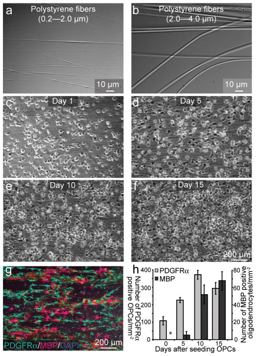
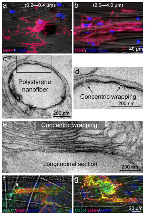
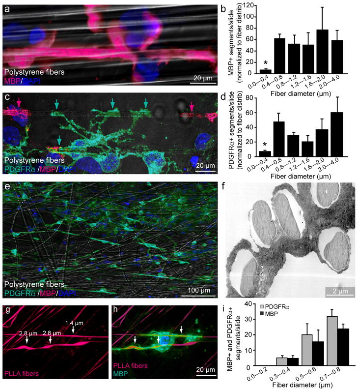
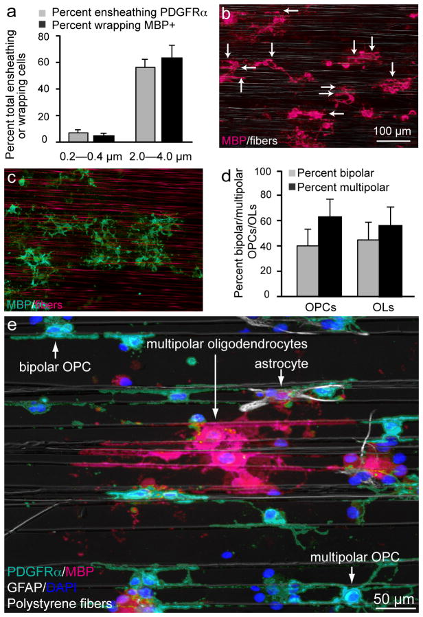
Current methods for studying central nervous system myelination necessitate permissive axonal substrates conducive to myelin wrapping by oligodendrocytes. We have developed a neuron-free culture system in which electron-spun nanofibers of varying sizes substitute for axons as a substrate for oligodendrocyte myelination, thereby allowing manipulation of the biophysical elements of axonal-oligodendroglial interactions. To investigate axonal regulation of myelination, this system effectively uncouples the role of molecular (inductive) cues from that of biophysical properties of the axon. We use this method to uncover the causation and sufficiency of fiber diameter in the initiation of concentric wrapping by rat oligodendrocytes. We also show that oligodendrocyte precursor cells display sensitivity to the biophysical properties of fiber diameter and initiate membrane ensheathment before differentiation. The use of nanofiber scaffolds will enable screening for potential therapeutic agents that promote oligodendrocyte differentiation and myelination and will also provide valuable insight into the processes involved in remyelination.


Conflict of interest statement

Competing Financial Interests

The authors declare no competing financial interests.

Figures

Figure 1
Figure 1
Temporal and spatial behavior of OPCs on polystyrene fibers are similar to that of OPCs on live axons. (a–f) Phase contrast images of (a, b) polystyrene nanofibers that range in diameter from (a) 0.2–2.0 μm (b) 2.0–4.0μm. (c–f) Bright-field images of cultured OPCs in the presence of exogenous PDGF and polystyrene fibers (0.2–0.4 μm) over 15 days in vitro (DIV). (g) Immunostaining of 10 DIV cultures against the OPC marker PDGFRα and MBP, which labels mature oligodendrocytes. (h) Quantification of the number of PDGFRα+ OPCs and MBP+ oligodendrocytes per millimeter squared on nanofibers as a function of time (n = 3, mean + SD,). Error bars represent SD. Scale bars, 10 μm (a, b), 200 μm (c–g).
Figure 2
Figure 2
Fiber diameter is sufficient to initiate myelination. (a,b) Immunostaining of cultures for MBP and DAPI in the presence of small (a, 0.2–0.4 μm) and large (b, 2.0–4.0 μm) diameter fibers. (c–e) Electron micrographs of an oligodendrocyte wrapping a large diameter nanofiber. d) Higher magnification of the inset box found in (c). Arrows indicate concentric wrapping of membrane. (e) A high magnification image of a longitudinal section illustrating an oligodendrocyte wrapping multiple layers of membrane around a large diameter nanofiber. (f, g) Immunostaining of oligodendrocytes cultured on large diameter nanofibers against the myelin proteins Myelin-Associated Glycoprotein (MAG) and Myelin Oligodendrocyte Glycoprotein (MOG). Scale bars, 40 μm (a, b), 200 nm (c–e), 20 μm (f, g).
Figure 3
Figure 3
Quantification of fiber diameter threshold and size preference for ensheathment and wrapping. (a) Immunostaining showing a magnified view of an MBP+ segment formed around a large diameter fiber (2.0–4.0 μm). Cell nuclei are labeled with DAPI (b) Quantification of the number of MBP+ segments normalized to the distribution of fiber diameters (n = 3, mean ± SEM.). (* represents P < 0.001, Tukey post hoc comparison after one-way ANOVA). (c) Immunostaining for PDGFRα and MBP illustrates an PDGFRα+/MBP− OPC in green ensheathing multiple large diameter fibers, as indicated by arrows. PDGFRα−/MBP+ oligodendrocyte segments in magenta (indicated by magenta arrows) are adjacent to the PDGFRα+/MBP− ensheathment. (d) Quantification of PDGFRα+ segments normalized to fiber diameter distribution (n = 3, mean ± SEM)(* represents P < 0.001, Tukey post hoc comparison after one-way ANOVA) (e) Low magnification image of OPC-fiber cultures immunostained at 5 DIV for PDGRα, MBP and DAPI. (f) Electron micrograph of OPC-fiber cultures illustrating ensheathment of multiple large diameter fibers. (g) Image of fibers engineered to display a range of diameter sizes continuously along the same fiber and conjugated with rhodamine. (h) Immunostaining for MBP illustrates an oligodendrocyte wrapping regions of the fiber that are larger in diameter as indicated by arrows. (i) Quantification of fiber diameter preference by binning the number of MBP+ and PDGFRα+ segments in 0.2 μm intervals from 0 to 0.8 μm (n =3, mean ± SEM). (P < 0.001, Tukey post hoc comparison after one-way ANOVA). Scale bars, 20 μm (a, c, h), 100 μm (e), 2 μm (f).
Figure 4
Figure 4
The majority of the cells in the cultures are capable of ensheathing and wrapping fibers above a minimum threshold. (a) Quantification of the percent of OPCs that ensheat or wrap small or large diameter fibers (n = 5, mean ± SD). Scale bar, 50 μm (b, c) Low magnification images of purified oligodendroglial-fiber cultures immunostained at 10 DIV for MBP. (b) Image of cultures in the presence of large (2.0–4.0 μm) diameter fibers. Fibers can be seen by phase contrast. Arrows indicate individual myelin-like segments. (c) Image of cultures in the presence of small diameter fibers (0.2–0.4 μm). As it is difficult to visualize small diameter fibers by phase contrast microscopy, they are labeled with rhodamine to allow for clear visualization. (d) Quantification of the differing morphologies of the oligodendroglial cells cultured on the nanofibers (n = 4, mean ± SD). Scale bars, 100 μm. (e) Low magnification image of purified oligodendroglial-fiber cultures immunostained at 10 DIV stained for glial fibrillary acidic protein (GFAP;white), a marker for contaminating astrocytes, PDFGRα (turquois), MBP (magenta), and DAPI (blue). Fibers can be seen by phase contrast. Arrows indicate each cell type and different morphologies.

Comment in

References

    1. Colello RJ, Pott U. Signals that initiate myelination in the developing mammalian nervous system. Mol Neurobiol. 1997;15:83–100. - PubMed
    1. Fruttiger M, Calver AR, Richardson WD. Platelet-derived growth factor is constitutively secreted from neuronal cell bodies but not from axons. Curr Biol. 2000;10:1283–1286. - PubMed
    1. Jiang F, Frederick TJ, Wood TL. IGF-I synergizes with FGF-2 to stimulate oligodendrocyte progenitor entry into the cell cycle. Dev Biol. 2001;232:414–423. - PubMed
    1. Nave KA, Trapp BD. Axon-glial signaling and the glial support of axon function. Annu Rev Neurosci. 2008;31:535–561. - PubMed
    1. Nave KA. Myelination and support of axonal integrity by glia. Nature. 2010;468:244–252. - PubMed

Publication types