. 2012 Nov;57(5):1044-51.
doi: 10.1016/j.jhep.2012.07.008. Epub 2012 Jul 14.

CXCR3-dependent recruitment and CCR6-mediated positioning of Th-17 cells in the inflamed liver

Affiliations

CXCR3-dependent recruitment and CCR6-mediated positioning of Th-17 cells in the inflamed liver

Ye Htun Oo et al. J Hepatol. 2012 Nov.

Abstract

Background & aims: IL-17 secreting CD4 (Th17) and CD8 (Tc17) T cells have been implicated in immune-mediated liver diseases, but the molecular basis for their recruitment and positioning within the liver is unknown.

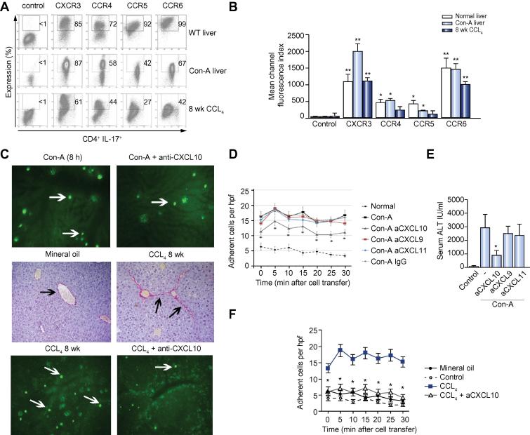
Methods: The phenotype and migratory behaviour of human liver-derived Th17 and Tc17 cells were investigated by flow cytometry and chemotaxis and flow-based adhesion assays. The recruitment of murine Th17 cells to the liver was studied in vivo using intra-vital microscopy.

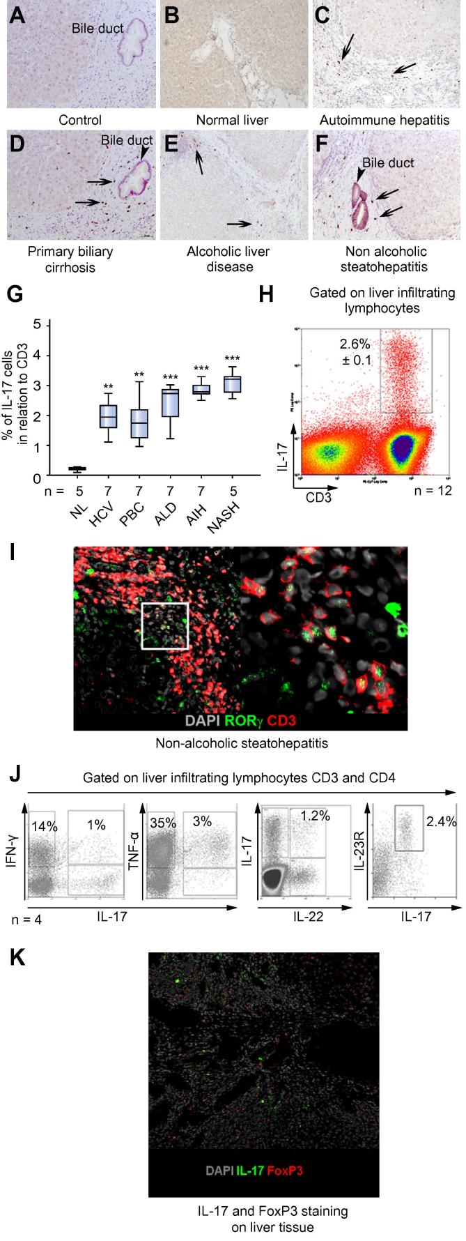
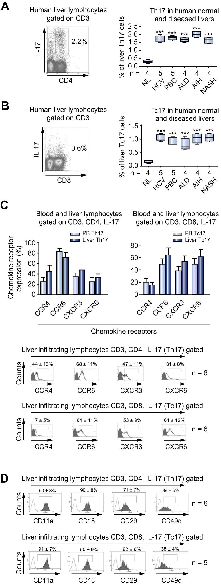
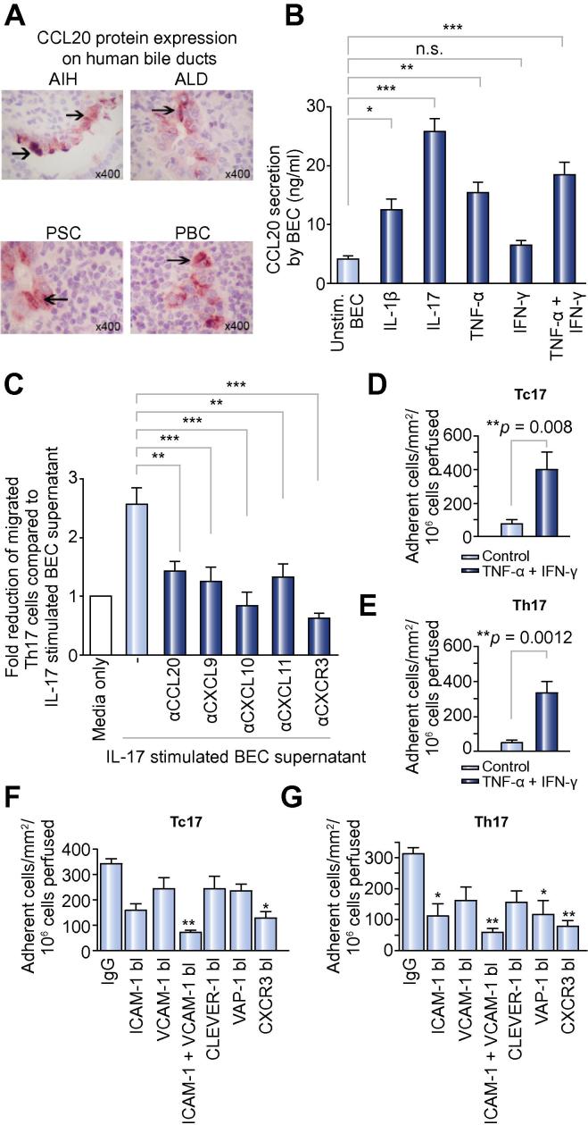
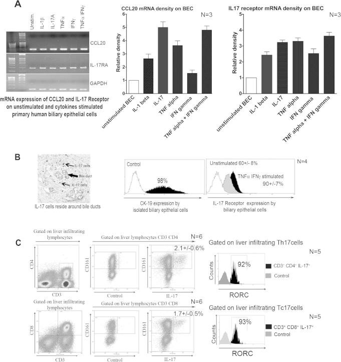
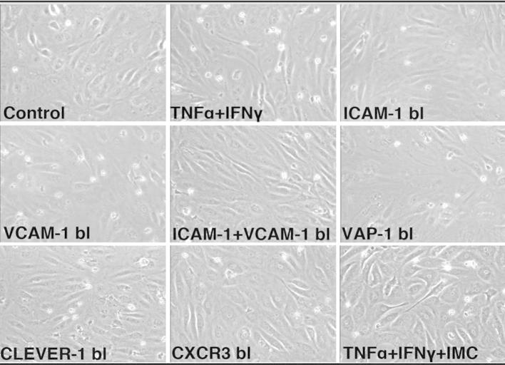
Results: IL-17(+) T cells comprised 1-3% of the T cell infiltrate in inflammatory liver diseases and included both CD4 (Th17) and CD8 (Tc17) cells. They expressed RORC and the IL-23 receptor and included subsets that secreted IL-22 and interferon-γ. Th17 and Tc17 cells expressed high levels of CXCR3 and CCR6, Tc17 cells also expressed CXCR6. Binding to human sinusoidal endothelium from flow was dependent on β1 and β2 integrins, CXCR3, and, in the case of Th17 cells, VAP-1. Th17 recruitment via sinusoids in mice with liver inflammation was reduced by treatment with antibodies against CXCR3 ligands, confirming the role of CXCR3 in Th17 recruitment in vivo. In human liver, IL-17(+) cells were detected in portal infiltrates close to inflamed bile ducts expressing the CCR6 ligand CCL20. Cytokine-treated human cholangiocytes secreted CCL20 and induced CCR6-dependent migration of Th17 cells suggesting that local cholangiocyte chemokine secretion localises Th17 cells to bile ducts.

Conclusions: CXCR3 promotes recruitment of Th17 cells from the blood into the liver in both human and murine liver injury. Their subsequent positioning near bile ducts is dependent on cholangiocyte-secreted CCL20.


Figures

Fig. 1
Fig. 1
Distribution and frequency of IL-17 producing cells in human liver. (A–F) IL-17+ cells (arrows) in human livers are shown. (A) Control antibody staining and IL-17A on (B) normal and (C–F) human livers from different diseases. Bile ducts are indicated by arrow-heads. (G) Proportion of CD3 T cells staining for IL-17 for different liver diseases analysed by immunohistochemistry counting (one-way ANOVA test; ∗∗∗⩽ 0.0001; ∗∗⩽ 0.001). (H) IL-17 expressing CD3 lymphocytes in human LIL (representative histogram of patient with autoimmune hepatitis, mean ± SEM; N = 12). (I) Confocal microscopy staining of RORc+ CD3+ cells in portal tracts; CD3 (red/TritC); RORc (green/FITC); nucleus (DAPI), magnification 10× (one representative sample of non-alcoholic steatohepatitis is shown; N = 4). The right hand panel shows an enlarged picture of marked area, magnification 40×. (J and Table 1) Percentage co-expression of different cytokines on liver infiltrating Th17 cells is shown. Liver infiltrating lymphocytes were stimulated for 5 h with PMA and ionomycin and brefeldin A Golgi block was applied for the last 2 h before staining for intracellular cytokines. LITh17 (liver infiltrating Th17) cells express the IL-23 receptor (liver infiltrating lymphocytes were gated on CD3 and CD4; representative flow plots of an ALD patient are shown). Four diseased livers were studied (2× AIH, PBC, and ALD). (K) Confocal images of FoxP3+ regulatory T cells and IL17 cells in a patient with PBC, FoxP3 (red/TritC), IL-17 (green/FITC), nucleus (DAPI). An autoimmune hepatitis liver is shown, magnification 10×. Data is representative of 4 samples. [This figure appears in colour on the web.]
Fig. 2
Fig. 2
Frequency and chemokine receptors expression on human intra-hepatic Th17 and Tc17 cells. (A) Liver infiltrating Th17 and (B) Tc17. One representative FACS blot of cells isolated from an autoimmune hepatitis liver is shown. Freshly isolated liver-infiltrating lymphocytes were gated on forward and side scatter and then re-gated on CD3. Percentage expression of Th17 and Tc17 in normal and diseased livers analysed by flow cytometry is shown (one-way ANOVA with Bonferroni post hoc correction; ∗∗∗⩽0.0001). (C) Percentage chemokine receptor expression on peripheral blood (N = 5) and liver-infiltrating Th17 and Tc17 cells (N = 6) is shown. Percentage expression is shown as mean ± SD. Bottom panel, flow cytometry overlay of the expression of liver infiltrating Th17 and Tc17 chemokine receptors (N = 6). (D) β1 and β2 integrin expression on Th17 and Tc17 by flow cytometry (representative overlay plot N = 6 for Th17; N = 5 for Tc17; percentage expression is shown as mean ± SD). [This figure appears in colour on the web.]
Fig. 3
Fig. 3
CCR6-dependent positioning around bile ducts and CXCR3-mediated recruitment of Th17 and Tc17. (A) CCL20 staining (arrow heads) of bile ducts on paraffin-embedded diseased human liver sections (AIH, autoimmune hepatitis; ALD, alcoholic liver disease; PSC, primary sclerosing cholangitis; PBC, primary biliary cirrhosis). (B) CCL20 secretion in human BEC supernatant determined by sandwich ELISA (one-way ANOVA test; ∗∗∗⩽0.0001; ∗∗⩽0.001; ⩽0.01; n.s., not significant) comparing medium alone to individual cytokine-stimulated BEC supernatant; N = 8 different diseased BEC. (C) Culture supernatants of IL-17-stimulated BEC were placed in the lower chambers for transwell assay and migration of Th17 quantified. Antibodies were used to deplete CCL20 (CCR6 ligand) and CXCL9–11 (CXCR3 ligands) in the BEC conditioned medium (N = 8, one-way ANOVA with Bonferroni correction, ∗∗<0.001; ∗∗∗<0.0001 comparing IL-17-stimulated BEC supernatant and supernatant after chemokine depletion/blockade or anti-CXCR3 treatment of Th17 cells). (D and E) Tc17 and Th17 lymphocyte adhesion to hepatic sinusoidal endothelial cells from flow. HSEC were stimulated with TNF-α (10 ng/ml) and IFN-γ (10 ng/ml) for 24 h prior to perfusion of (D) Tc17 and (E) Th17 lymphocytes at a shear stress of 0.05 Pa. Adhesion was classified as rolling, static adhesion or migration, which were combined to give the total number of adherent cells ± SEM from N = 4 different Tc17 and Th17 cell preparations (F and G). Adhesion of Th17 and Tc17 cells (total numbers) on TNF-α/IFN-γ-stimulated HSEC was reduced by function-blocking antibodies against ICAM-1 and VCAM-1, CLEVER-1, VAP-1, on HSEC or anti-CXCR3 block on Tc17/Th17 cells compared to adhesion observed with a control antibody (F and G, Supplementary Fig. 2). <0.01, ∗∗<0.001 by one-way ANOVA with Bonferroni correction. [This figure appears in colour on the web.]
Fig. 4
Fig. 4
Chemokine-mediated Th17 recruitment to the injured murine liver. (A and B) Chemokine receptor expression by flow cytometry on liver-derived Th17 cells from ConA-induced acute hepatitis and CCL4-induced chronic liver injury (mean ± SEM. N = 6; <0.05; ∗∗<0.01; Paired t test). (C, D and F) Ex-vivo generated, CFSE-labelled Th17 were injected (C and D) 8 h after ConA or (C and F) 1 week after week 8 CCL4 injection, and recruitment investigated by IVM. Still images of treatment adherent cells in hepatic sinusoids after 30 min are shown (C, D and F). The graph shows the mean number of adherent cells from six high-power fields ± SEM at each time point with at least 3 animals/group for (D) ConA and (F) CCL4. Data are compared to control animals that received saline injections. Anti-CXCL9–11 or control antibodies were injected 1 h after (D) ConA injection and (F) anti-CXCL10 or control antibodies for CCL4. (C, middle panel) CCL4 liver fibrosis was confirmed by Sirius Red staining (black arrows). (E) Serum ALT levels after ConA injection comparing blocking CXCL9–11. (Data represents mean ± SEM. N = 6; <0.05; paired t test.) [This figure appears in colour on the web.]
Supplementary Fig. 1
Supplementary Fig. 2

Comment in

Similar articles

Cited by

References

    1. Harrington L.E., Hatton R.D., Mangan P.R., Turner H., Murphy T.L., Murphy K.M., et al. Interleukin 17-producing CD4+ effector T cells develop via a lineage distinct from the T helper type 1 and 2 lineages. Nat Immunol. 2005;6:1123–1132. - PubMed
    1. Korn T., Bettelli E., Oukka M., Kuchroo V.K. IL-17 and Th17 cells. Annu Rev Immunol. 2009;27:485–517. - PubMed
    1. Ivanov I.I., McKenzie B.S., Zhou L., Tadokoro C.E., Lepelley A., Lafaille J.J., et al. The orphan nuclear receptor RORgammat directs the differentiation program of proinflammatory IL-17+ T helper cells. Cell. 2006;126:1121–1133. - PubMed
    1. Unutmaz D. RORC2: the master of human Th17 cell programming. Eur J Immunol. 2009;39:1452–1455. - PubMed
    1. Veldhoen M., Uyttenhove C., van S.J., Helmby H., Westendorf A., Buer J., et al. Transforming growth factor-beta ‘reprograms’ the differentiation of T helper 2 cells and promotes an interleukin 9-producing subset. Nat Immunol. 2008;9:1341–1346. - PubMed

Publication types