Review
. 2012 Jul-Aug;6(4):374-84.
doi: 10.4161/cam.20832. Epub 2012 Jul 1.

L1CAM: a major driver for tumor cell invasion and motility

Affiliations
Review

L1CAM: a major driver for tumor cell invasion and motility

Helena Kiefel et al. Cell Adh Migr. 2012 Jul-Aug.

Abstract

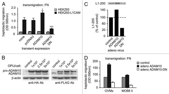
The L1 cell adhesion molecule (L1CAM) plays a major role in the development of the nervous system and in the malignancy of human tumors. In terms of biological function, L1CAM comes along in two different flavors: (1) a static function as a cell adhesion molecule that acts as a glue between cells; (2) a motility promoting function that drives cell migration during neural development and supports metastasis of human cancers. Important factors that contribute to the switch in the functional mode of L1CAM are: (1) the cleavage from the cell surface by membrane proximal proteolysis and (2) the ability to change binding partners and engage in L1CAM-integrin binding. Recent studies have shown that the cleavage of L1CAM by metalloproteinases and the binding of L1CAM to integrins via its RGD-motif in the sixth Ig-domain activate signaling pathways distinct from the ones elicited by homophilic binding. Here we highlight important features of L1CAM proteolysis and the signaling of L1CAM via integrin engagement. The novel insights into L1CAM downstream signaling and its regulation during tumor progression and epithelial-mesenchymal transition (EMT) will lead to a better understanding of the dualistic role of L1CAM as a cell adhesion and/or motility promoting cell surface molecule.


Figures

None
Figure 1. L1CAM structure and cleavage. (A) L1CAM is a type I transmembrane molecule of the immunoglobulin superfamily. It is composed of sixth immunoglobulin domains, five fibronectin-type III repeats and a conserved cytoplasmic domain. L1CAM can bind homophilically to other L1CAM molecules or heterophilically to various ligands. The RGD-site in the sixth Ig-domain supports binding to integrins such as α5β1 αvβ3 or αvβ5. L1CAM can be cleaved proximal to the plasma membrane by the metalloproteinases ADAM10 and ADAM17. This ectodomain shedding results in the release of the 200 kDa soluble ectodomain whereas the 32 kDa transmembrane stub is retained in the plasma membrane (see illustration). The RGD motif and the sites of proteolytic cleavage are specified. (B) The membrane proximal cleavage site. The amino acid sequence of mouse and human L1CAM is shown and the putative site of ADAM-mediated cleavage is indicated. It should be noted that the L1-32 cleavage fragment is insensitive to Endoglycosidase F treatment suggesting that it is devoid of N-linked glycans.
None
Figure 2. Proteolytic cleavage and nuclear translocation of L1CAM-ICD. Following ADAM-mediated ectodomain shedding, the membraneous 32 kDa stub of L1CAM functions as a substrate for the intramembraneous cleavage by γ-secretase/presenilin. The resulting fragment represents the intracellular domain of L1CAM (L1-ICD). L1-ICD can translocate into the nucleus by an unknown mechanism and participate in the transcriptional regulation of genes including Cathepsin B, CRABPII, β3-integrin and MDK.
None
Figure 3. L1CAM and its ADAM-mediated cleavage promote cell motility. (A) Stable overexpression of L1CAM in HEK293 cells promotes haptotactic cell migration in a Boyden chamber migration assay with fibronectin (FN) as substrate. Transient overexpression of ADAM10 augments, whereas a dominant-negative, proteolytically non-active form of ADAM10 (ADAM10-DN) blocks cell migration. (B) Adenovirus encoding ADAM10 (HA-tagged) or ADAM10-DN (FLAG tagged) were transduced in OVMz cells (L1CAM positive) and cell lysated were probed with the indicated mAbs to detect successful overexpression. (C) Detection of soluble L1CAM in the culture supernatant of transduced cells. The supernantant from equal number of cells was depleted from cellular debris by centrifugation and soluble proteins were precipitated by the addition of TCA (trichloroacetic acid). Soluble L1-200 was detected after SDS-PAGE and western blot analysis as described. (D) Haptotactic migration of ovarian carcinoma cell lines OVMz and MO68II (both L1CAM positive) after transduction with adenovirus.
None
Figure 4. L1CAM can trigger different signaling pathways. Schematic illustration of distinct signaling pathways triggered by L1CAM due to the interacting with different binding partners. (A) L1CAM homophilic interactions promote static cell-cell binding and trigger predominantly the MAPK pathway. This signaling can be modulated by interactions with growth factor receptors (GFR). (B) The binding of L1CAM to integrins triggers NFκB activation and renders cells more motile and invasive. Cleavage of L1CAM from the membrane generates soluble L1CAM. The intracellular fragment L1-ICD can translocate into the nucleus and activate gene transcription. (C) Upregulation of L1CAM by TGF-β allows L1CAM-integrin downstream signaling via FAK-Src. This pathway induces production and release of IL-1β that in turn activates NFκB via binding to the IL-1 receptor (IL-1RI).
None
Figure 5. L1CAM upregulation during EMT. Carcinoma cells at the primary tumor site can lose their epithelial phenotype (charcterized by expression of E-cadherin and keratins) and undergo EMT-like phenotypic changes under the influence of TGF-β produced by fibroblasts in the tumor stroma. Tumor cells possess a more mesenchymal phenotype charcterized by upregulation of vimentin, SLUG and L1CAM. L1CAM at the tumor invasive front is eventually cleaved by ADAMs producing soluble L1CAM. By interacting with integrins on neighboring tumor cells, stroma cells or invaded leukocytes, L1CAM induces NFkB activity via IL-1β or other factors. L1CAM expression thereby promotes cell motility and invasion.

References

    1. Arlt A, Vorndamm J, Müerköster S, Yu H, Schmidt WE, Fölsch UR, et al. Autocrine production of interleukin 1beta confers constitutive nuclear factor kappaB activity and chemoresistance in pancreatic carcinoma cell lines. Cancer Res. 2002;62:910–6. - PubMed
    1. Moos M, Tacke R, Scherer H, Teplow D, Früh K, Schachner M. Neural adhesion molecule L1 as a member of the immunoglobulin superfamily with binding domains similar to fibronectin. Nature. 1988;334:701–3. doi: 10.1038/334701a0. - DOI - PubMed
    1. Maness PF, Schachner M. Neural recognition molecules of the immunoglobulin superfamily: signaling transducers of axon guidance and neuronal migration. Nat Neurosci. 2007;10:19–26. doi: 10.1038/nn1827. - DOI - PubMed
    1. Rathjen FG, Schachner M. Immunocytological and biochemical characterization of a new neuronal cell surface component (L1 antigen) which is involved in cell adhesion. EMBO J. 1984;3:1–10. - PMC - PubMed
    1. Wolff JM, Frank R, Mujoo K, Spiro RC, Reisfeld RA, Rathjen FG. A human brain glycoprotein related to the mouse cell adhesion molecule L1. J Biol Chem. 1988;263:11943–7. - PubMed

Publication types

Substances

LinkOut - more resources