. 2012 Sep;23(9):1467-73.
doi: 10.1681/ASN.2011100967. Epub 2012 Jul 12.

Cellular senescence limits regenerative capacity and allograft survival

Affiliations

Cellular senescence limits regenerative capacity and allograft survival

Heidi Braun et al. J Am Soc Nephrol. 2012 Sep.

Abstract

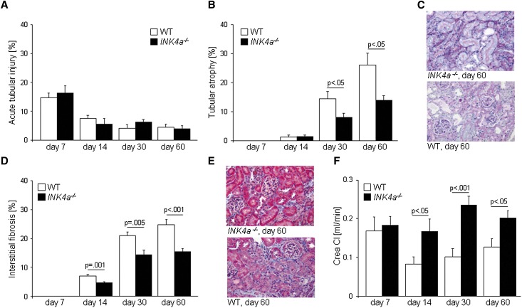
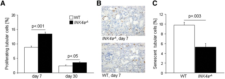
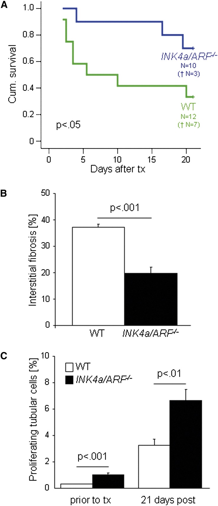
Long-term graft survival after kidney transplantation remains unsatisfactory and unpredictable. Interstitial fibrosis and tubular atrophy are major contributors to late graft loss; features of tubular cell senescence, such as increased p16(INK4a) expression, associate with these tubulointerstitial changes, but it is unknown whether the relationship is causal. Here, loss of the INK4a locus in mice, which allows escape from p16(INK4a)-dependent senescence, significantly reduced interstitial fibrosis and tubular atrophy and associated with improved renal function, conservation of nephron mass, and transplant survival. Compared with wild-type controls, kidneys from INK4a(-/-) mice developed significantly less interstitial fibrosis and tubular atrophy after ischemia-reperfusion injury. Consistently, mice that received kidney transplants from INK4a/ARF(-/-) donors had significantly better survival 21 days after life-supporting kidney transplantation and developed less tubulointerstitial changes. This correlated with higher proliferative rates of tubular cells and significantly fewer senescent cells. Taken together, these data suggest a pathogenic role of renal cellular senescence in the development of interstitial fibrosis and tubular atrophy and kidney graft deterioration by preventing the recovery from injury. Inhibiting premature senescence could have therapeutic benefit in kidney transplantation but has to be balanced against the risks of suspending antitumor defenses.


Figures

Figure 1.
Figure 1.
Ischemia-reperfusion injury in INK4a−/− and wild-type mice. (A) Acute tubular injury (shown as percentage of affected area) is most pronounced 7 days after injury and decreases thereafter. There are no differences between the groups. (B) Tubular atrophy (percentage of affected area) is first detected 14 days after injury. Significant differences occur 30 and 60 days after injury, with INK4a−/− kidneys developing less tubular atrophy. (C) Representative sections from kidneys 60 days after injury. Tubular atrophy is visualized with the periodic acid–Schiff staining. (D) Interstitial fibrosis (percentage of blue-stained area) reveals a similar picture to tubular atrophy with significantly less interstitial fibrosis seen in INK4a−/− kidneys 14, 30, and 60 days after injury. (E) Representative Masson Trichrome staining of kidneys 60 days after injury. Interstitial fibrosis is visualized as blue-colored collagen fibers. (F) Creatinine clearances are significantly lower in wild-type mice 14, 30, and 60 days after injury. WT, wild-type; Crea Cl, creatine clearance. Original magnification, ×200.
Figure 2.
Figure 2.
Proliferation and senescence of tubular cells from INK4a−/− and wild-type kidneys after ischemia-reperfusion injury. (A) Tubular cell proliferation (percentage of Ki67 positive tubular cells) measured 7 and 30 days after injury was significantly higher in kidneys from INK4a−/− mice. Proliferation rates declined from day 7 to day 30 in both groups (P<0.05). (B) Representative staining showing Ki67-positive cells in kidneys 7 days after injury. (C) The number of senescent tubular cells at day 60 after injury was significantly lower in kidneys from INK4a−/− mice. WT, wild-type. Original magnification, ×200.
Figure 3.
Figure 3.
Life-supporting allogeneic renal transplantation of INK4a/ARF−/− and wild-type kidneys. (A) Cumulative survival was significantly better in mice that received a transplant from a INK4a/ARF−/− mouse. (B) Interstitial fibrosis (percentage of blue-stained area) was significantly higher in wild-type transplants. (C) Tubular cell proliferation (percentage of Ki67-positive tubular cells) was significantly higher in INK4a/ARF−/− transplants, before and 21 days after transplantation. WT, wild-type; tx, transplant.

References

    1. Lamb KE, Lodhi S, Meier-Kriesche HU: Long-term renal allograft survival in the United States: A critical reappraisal. Am J Transplant 11: 450–462, 2011 - PubMed
    1. Einecke G, Sis B, Reeve J, Mengel M, Campbell PM, Hidalgo LG, Kaplan B, Halloran PF: Antibody-mediated microcirculation injury is the major cause of late kidney transplant failure. Am J Transplant 9: 2520–2531, 2009 - PubMed
    1. Nath KA: Tubulointerstitial changes as a major determinant in the progression of renal damage. Am J Kidney Dis 20: 1–17, 1992 - PubMed
    1. Nankivell BJ, Borrows RJ, Fung CL, O’Connell PJ, Allen RD, Chapman JR: The natural history of chronic allograft nephropathy. N Engl J Med 349: 2326–2333, 2003 - PubMed
    1. Nakorchevsky A, Hewel JA, Kurian SM, Mondala TS, Campbell D, Head SR, Marsh CL, Yates JR, 3rd, Salomon DR: Molecular mechanisms of chronic kidney transplant rejection via large-scale proteogenomic analysis of tissue biopsies. J Am Soc Nephrol 21: 362–373, 2010 - PMC - PubMed

Publication types

MeSH terms

Substances