. 2012 Aug;23(8):1339-50.
doi: 10.1681/ASN.2012010017. Epub 2012 Jul 12.

Loss of the endothelial glycocalyx links albuminuria and vascular dysfunction

Affiliations

Loss of the endothelial glycocalyx links albuminuria and vascular dysfunction

Andrew H J Salmon et al. J Am Soc Nephrol. 2012 Aug.

Abstract

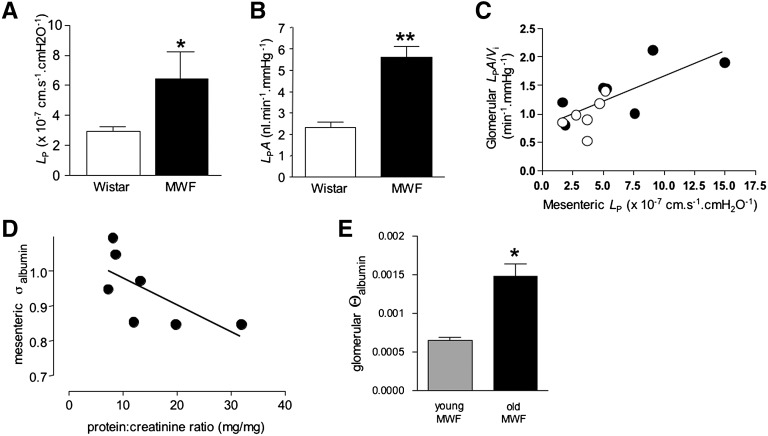
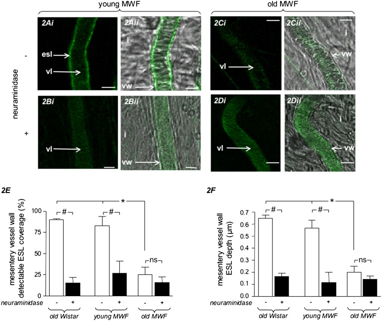
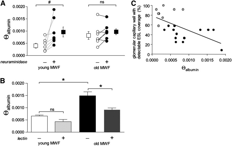
Patients with albuminuria and CKD frequently have vascular dysfunction but the underlying mechanisms remain unclear. Because the endothelial surface layer, a meshwork of surface-bound and loosely adherent glycosaminoglycans and proteoglycans, modulates vascular function, its loss could contribute to both renal and systemic vascular dysfunction in proteinuric CKD. Using Munich-Wistar-Fromter (MWF) rats as a model of spontaneous albuminuric CKD, multiphoton fluorescence imaging and single-vessel physiology measurements revealed that old MWF rats exhibited widespread loss of the endothelial surface layer in parallel with defects in microvascular permeability to both water and albumin, in both continuous mesenteric microvessels and fenestrated glomerular microvessels. In contrast to young MWF rats, enzymatic disruption of the endothelial surface layer in old MWF rats resulted in neither additional loss of the layer nor additional changes in permeability. Intravenous injection of wheat germ agglutinin lectin and its adsorption onto the endothelial surface layer significantly improved glomerular albumin permeability. Taken together, these results suggest that widespread loss of the endothelial surface layer links albuminuric kidney disease with systemic vascular dysfunction, providing a potential therapeutic target for proteinuric kidney disease.


Figures

Figure 1.
Figure 1.
Widespread increase in microvascular permeability in old MWF rats with proteinuric kidney disease. (A) Mean (±SEM) hydraulic conductivity (LP) of mesenteric microvessels was significantly elevated in MWF rats (black bar; n=7 animals and vessels), compared with healthy Wistar rats (open bar; n=15 animals and vessels). (B) Mean (±SEM) glomerular Kf, normalized for glomerular volume (LPA/Vi) was significantly elevated in glomeruli from MWF rats (black bar; n=58 glomeruli from 10 animals) compared with glomeruli isolated from healthy Wistar rats (open bar; n=24 glomeruli from 6 animals). (C) Glomerular LPA/Vi and mesenteric LP values from the same animal were positively correlated (Pearson r=0.58; P<0.05) (open circles, Wistar rats; filled circles, MWF rats). (D) There was a significant, negative relation between mesenteric microvessel reflection coefficient (mesenteric σ) and urinary protein/creatinine ratio in MWF rats (Spearman r=−0.71; P<0.05). (E) Glomerular albumin sieving coefficient (Θalbumin) was low in healthy young MWF rats (n=11 glomeruli in 3 animals), and significantly elevated in old MWF rats with proteinuric kidney disease (n=20 glomeruli in 5 animals). *P<0.05 (unpaired t test). **P<0.01 (unpaired t test).
Figure 2.
Figure 2.
ESL of mesenteric microvessels is compromised in old MWF rats. Confocal microscopy images of FITC-WGA lectin (green) labeling ESL (esl), alongside simultaneous DIC imaging of microvessel anatomy in a healthy young MWF rat (A and B) and an old MWF rat (C and D). Linear labeling of ESL at the vessel margin in a young MWF rat (Ai and Aii) is removed by neuraminidase (Bi and Bii). ESL is not apparent in mesenteric microvessels of old MWF rats before (Ci and Cii) or after (Di and Dii) neuraminidase. Under baseline conditions (open bars), ESL covers most of wall of mesenteric microvessels (E), and is approximately 0.6 µm deep (F), in healthy rats (old Wistar rats, n=111 vessels in 3 rats; young MWF rats, n=47 vessels in 3 rats). ESL coverage and depth are both reduced by neuraminidase (black bar) in these healthy rats. In contrast, ESL is significantly sparser (E) and shallower (F) in old MWF rats under baseline conditions (n=165 vessels in 4 rats), and is not reduced any further by neuraminidase. *P<0.05 versus both healthy rat groups (one-way ANOVA, Bonferroni); #P<0.05 versus baseline values (one-way ANOVA, Bonferroni); ns, not significant versus preneuraminidase values (one-way ANOVA, Bonferroni). esl, ESL; vl, vessel lumen; i, interstitium; vw, vessel wall. Scale bar, 10 microns.
Figure 3.
Figure 3.
ESL of glomerular capillaries is compromised in older MWF rats. (A) Under baseline conditions, multiphoton microscopy imaging reveals a linear label (white arrows) of FITC-WGA lectin (green) at the margin of superficial glomerular capillaries containing fluorescently labeled albumin (red). (B) Again under baseline conditions, linear labeling with the same WGA lectin (but tagged with the red fluorophore alexa-594) is on the luminal surface of lucifer yellow-labeled glomerular endothelial cells (green), confirming that the WGA lectin labels glomerular ESL. (Bii) High-magnification image of the rectangular region outlined in Bi. (C) After neuraminidase treatment, the majority of glomerular capillary wall sites do not exhibit linear glomerular ESL label (open arrowheads), although some linear label is still apparent (white arrows). (D) Imaging of the same optical section of the same glomerulus shown in C after neuraminidase treatment. Most of the ESL label has been removed as observed at low magnification (open arrowheads in Di) and high magnification (broken lines in Di). (E) Under baseline conditions (open circles), the majority of glomerular capillary wall sites in young MWF rats exhibit ESL labeling. Neuraminidase treatment of these healthy animals (black circles) significantly reduces glomerular ESL labeling. (F) In old MWF rats, multiphoton microscopy imaging of FITC-WGA lectin (green) reveals very little linear labeling (white arrow) at the margin of superficial glomerular capillaries containing fluorescently labeled albumin (red). The majority of glomerular capillary wall sites do not display linear labeling (open arrowhead). (G) After neuraminidase treatment of an old MWF rat, there is little change in the pattern of WGA lectin labeling (labeling of the brush border of tubular epithelium confirms the presence of detectable FITC-WGA lectin). (H) ESL coverage in old MWF rats under baseline conditions (open circles) is less than in young rats (compared with 3E), and is not further reduced by neuraminidase (closed circles. Each circle represents mean ± SEM values from 4 to 10 glomeruli in individual animals. $P<0.05 versus baseline (one-way ANOVA, Bonferroni); #P<0.05 (one-way ANOVA, Bonferroni); ns, P>0.05 versus baseline (one-way ANOVA, Bonferroni). gc, glomerular capillaries; ec, endothelial cell; esl, ESL; *, extracapsular capillary; pus, peripheral urinary space; P, podocyte (negatively labeled by lucifer yellow); t, tubular epithelium. Scale bar, 10 µm in A, C, F, and G; 5 µm in Bi, Bii, Di, and Dii.
Figure 4.
Figure 4.
Loss of glomerular ESL contributes to increased glomerular albumin sieving coefficient, and modification of ESL improves glomerular albumin sieving coefficient, in old MWF rats. (A) In healthy young MWF rats, glomerular albumin sieving coefficient values (Θalbumin) of individual glomeruli (open circles; open square represents mean ± SEM) were elevated after exposure to neuraminidase in every glomerulus (black circles; black square represents mean ± SEM). #P<0.05 versus baseline (paired t test). In contrast, higher Θalbumin values in old MWF rats (open circles; open square represents mean ± SEM) were not significantly elevated any further after neuraminidase exposure (black circles; black square represents mean ± SEM) ns, P>0.05 versus baseline (paired t test). (B) Binding of WGA lectin to the ESL (hashed bars) significantly reduced Θalbumin in old MWF rats (black bar). A similar, nonsignificant trend toward reduced Θalbumin after lectin binding was also observed in young MWF rats (open bar). *P<0.05; ns, P>0.05 versus baseline (one-way ANOVA, Bonferroni). (C) Θalbumin for individual glomeruli (gray circles, young MWF; black circles, old MWF) were significantly correlated with the percentage of glomerular capillary wall sites with ESL cover in the same glomerulus (Pearson r=−0.59; P<0.005).

References

    1. Matsushita K, van der Velde M, Astor BC, Woodward M, Levey AS, de Jong PE, Coresh J, Gansevoort RT, Chronic Kidney Disease Prognosis Consortium : Association of estimated glomerular filtration rate and albuminuria with all-cause and cardiovascular mortality in general population cohorts: A collaborative meta-analysis. Lancet 375: 2073–2081, 2010 - PMC - PubMed
    1. Hillege HL, Fidler V, Diercks GF, van Gilst WH, de Zeeuw D, van Veldhuisen DJ, Gans RO, Janssen WM, Grobbee DE, de Jong PE, Prevention of Renal and Vascular End Stage Disease (PREVEND) Study Group : Urinary albumin excretion predicts cardiovascular and noncardiovascular mortality in general population. Circulation 106: 1777–1782, 2002 - PubMed
    1. Tatematsu S, Wakino S, Kanda T, Homma K, Yoshioka K, Hasegawa K, Sugano N, Kimoto M, Saruta T, Hayashi K: Role of nitric oxide-producing and -degrading pathways in coronary endothelial dysfunction in chronic kidney disease. J Am Soc Nephrol 18: 741–749, 2007 - PubMed
    1. Clausen P, Jensen JS, Jensen G, Borch-Johnsen K, Feldt-Rasmussen B: Elevated urinary albumin excretion is associated with impaired arterial dilatory capacity in clinically healthy subjects. Circulation 103: 1869–1874, 2001 - PubMed
    1. Stehouwer CD, Henry RM, Dekker JM, Nijpels G, Heine RJ, Bouter LM: Microalbuminuria is associated with impaired brachial artery, flow-mediated vasodilation in elderly individuals without and with diabetes: Further evidence for a link between microalbuminuria and endothelial dysfunction—the Hoorn Study. Kidney Int Suppl 92: S42–S44, 2004 - PubMed

Publication types