

Genome mapping on nanochannel arrays for structural variation analysis and sequence assembly
- PMID: 22797562
- PMCID: PMC3817024
- DOI: 10.1038/nbt.2303
Genome mapping on nanochannel arrays for structural variation analysis and sequence assembly
Abstract
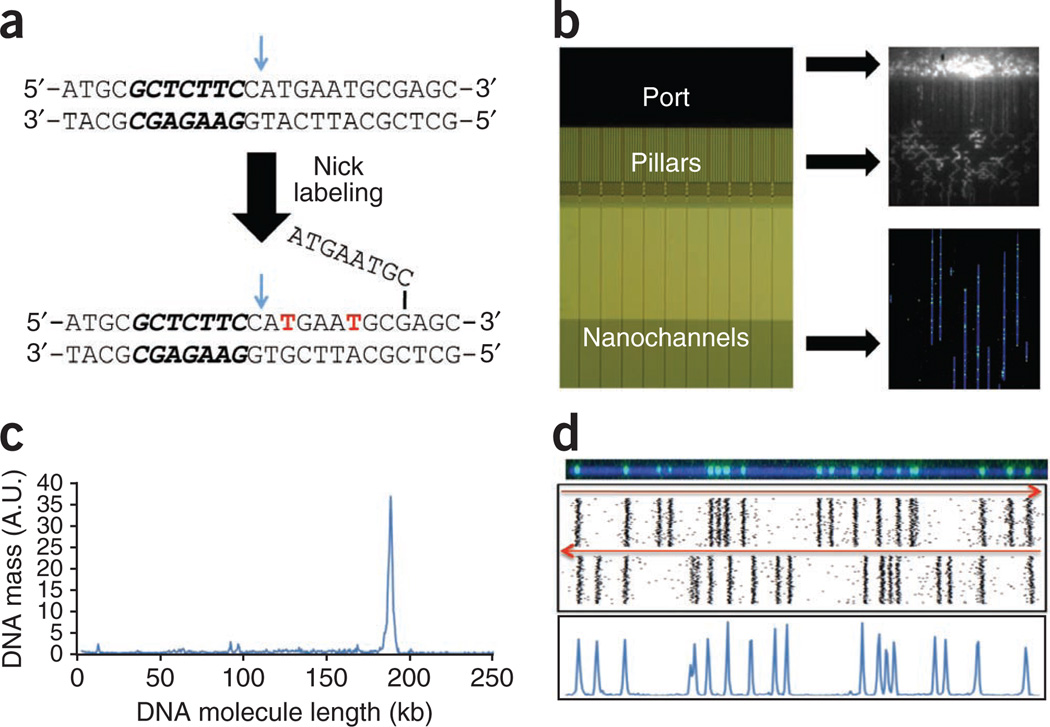
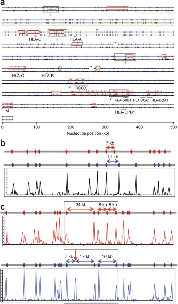
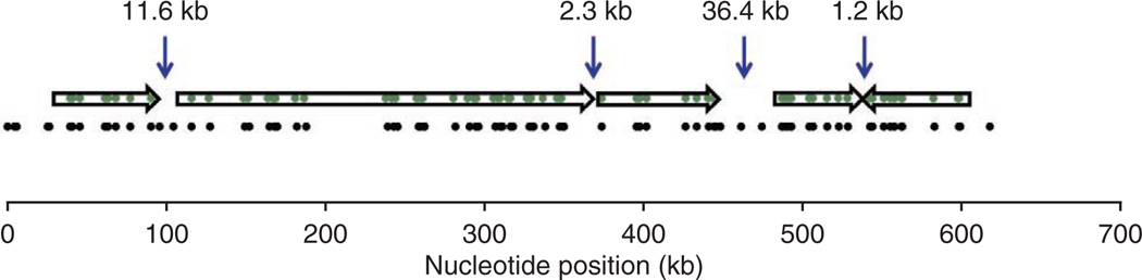
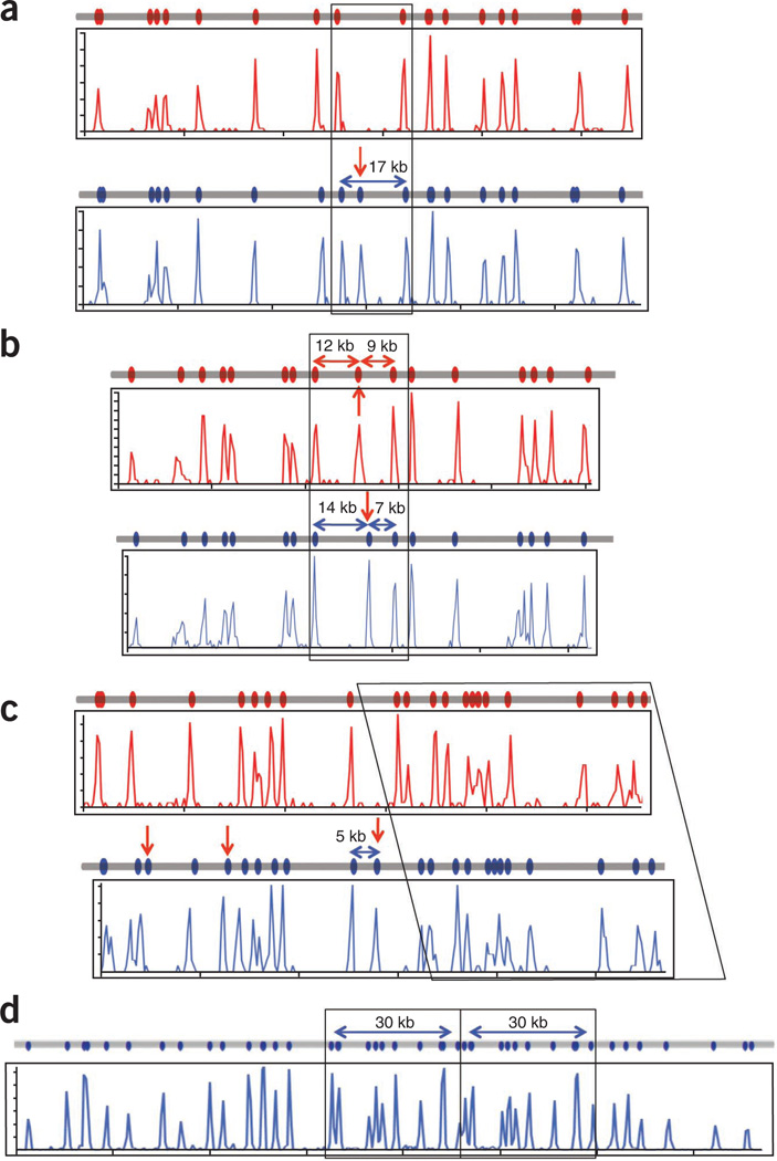
We describe genome mapping on nanochannel arrays. In this approach, specific sequence motifs in single DNA molecules are fluorescently labeled, and the DNA molecules are uniformly stretched in thousands of silicon channels on a nanofluidic device. Fluorescence imaging allows the construction of maps of the physical distances between occurrences of the sequence motifs. We demonstrate the analysis, individually and as mixtures, of 95 bacterial artificial chromosome (BAC) clones that cover the 4.7-Mb human major histocompatibility complex region. We obtain accurate, haplotype-resolved, sequence motif maps hundreds of kilobases in length, resulting in a median coverage of 114× for the BACs. The final sequence motif map assembly contains three contigs. With an average distance of 9 kb between labels, we detect 22 haplotype differences. We also use the sequence motif maps to provide scaffolds for de novo assembly of sequencing data. Nanochannel genome mapping should facilitate de novo assembly of sequencing reads from complex regions in diploid organisms, haplotype and structural variation analysis and comparative genomics.
Figures


Comment in
-
Channeling DNA for optical mapping.Nat Biotechnol. 2012 Aug;30(8):762-3. doi: 10.1038/nbt.2324. Nat Biotechnol. 2012. PMID: 22871713 No abstract available.
-
DNA: stretch for the camera.Nat Methods. 2012 Sep;9(9):862-3. doi: 10.1038/nmeth.2166. Nat Methods. 2012. PMID: 23097782 No abstract available.
References
Publication types
MeSH terms
Substances
Grants and funding
LinkOut - more resources
Full Text Sources
Other Literature Sources
Medical
Miscellaneous
