. 2012 Sep;8(9):759-68.
doi: 10.1038/nchembio.1021. Epub 2012 Jul 15.

Architectural and thermodynamic principles underlying intramembrane protease function

Affiliations

Architectural and thermodynamic principles underlying intramembrane protease function

Rosanna P Baker et al. Nat Chem Biol. 2012 Sep.

Abstract

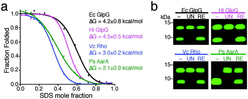
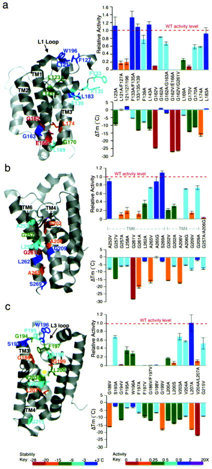
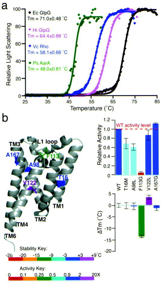
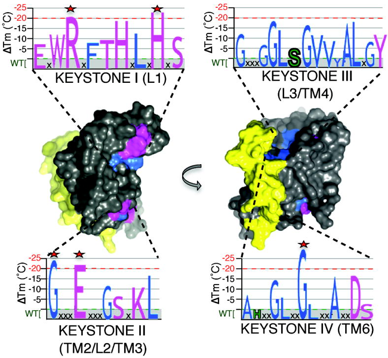
Intramembrane proteases hydrolyze peptide bonds within the membrane as a signaling paradigm universal to all life forms and with implications in disease. Deciphering the architectural strategies supporting intramembrane proteolysis is an essential but unattained goal. We integrated new, quantitative and high-throughput thermal light-scattering technology, reversible equilibrium unfolding and refolding and quantitative protease assays to interrogate rhomboid architecture with 151 purified variants. Rhomboid proteases maintain low intrinsic thermodynamic stability (ΔG = 2.1-4.5 kcal mol(-1)) resulting from a multitude of generally weak transmembrane packing interactions, making them highly responsive to their environment. Stability is consolidated by two buried glycines and several packing leucines, with a few multifaceted hydrogen bonds strategically deployed to two peripheral regions. Opposite these regions lie transmembrane segment 5 and connected loops that are notably exempt of structural responsibility, suggesting intramembrane proteolysis involves considerable but localized protein dynamics. Our analyses provide a comprehensive 'heat map' of the physiochemical anatomy underlying membrane-immersed enzyme function at, what is to our knowledge, unprecedented resolution.


Conflict of interest statement

COMPETING FINANCIAL INTERESTS STATEMENT

The authors declare no conflict of interest exists.

Figures

Figure 1
Figure 1. Thermodynamic assessment of rhomboid protease stability
(a) Chemical unfolding of wildtype rhomboid proteases in 2mM DDM at 25°C monitored by protease activity. The fraction folded was plotted against SDS mole fraction and the curves were fit with a two-state model according to the Santoro-Bolen equation. Ec is Escherichia coli, Vc is Vibrio cholerae, Hi is Haemophilus influenzae, and Ps is Providencia stuartii. (b) Activity analysis of refolded rhomboid proteases. ‘–’ denotes enzyme prior to treatment, ‘UN’ the fully-unfolded state (0.9 mole fraction SDS), and ‘RE’ the refolded state (by dilution from 0.9 to 0.1 mole fraction SDS). Shown are infrared fluorescence western analyses in which the top band is the substrate while the lower band is the cleaved product. Refolding restored 100% activity for all rhomboid proteases tested except AarA (~50%).
Figure 2
Figure 2. Differential static light scattering as a probe of rhomboid stability
(a) Static light scattering quantified every 0.5°C was plotted against temperature (°C) for E. coli GlpG. The transition midpoint (Tm) for wildtype GlpG was 71.0±0.48°C (n=24). Note that the red line denotes loss of protease activity (not light scattering) and the purple line monitors helicity (ellipticity at 222 nm) as a function of temperature. The lower panel shows a western analysis of GlpG protease activity after pretreatment at varying temperatures. (b) Thermal denaturation of GlpG is irreversible: black bars show protease activity (mean ± standard deviation) conducted at the designated temperatures for 20 minutes, while the blue bars denote GlpG preincubated at the designated temperatures for 20 minutes, cooled, and tested for activity at 37°C. (c) Effect of protein concentration and rate of heating on the Tm (± standard deviation) of wildtype GlpG. (d). Quantified change in Tm (ΔTm) of all engineered GlpG variants plotted against primary structure (residue numbers). Shown are substitutions to alanine and glycine-to-valine mutants in each residue. Colors and segment numbering correlate with tertiary structure (lateral view from membrane, cytoplasm down). The five most perturbed variants (R137A, H145A, G162V, E166A, G261V) are highlighted with red bars (ΔTm>-20°C).
Figure 3
Figure 3. Only two regions of hydrogen bonding are critical for GlpG stability
(a) The hydrogen bonds (in pink throughout) made by the TM2 residue E166 (inset) including to T97 and S171 make critical contributions to GlpG thermostability, while intramembrane hydrogen bonds Y138, W158, Y160, and T178 make only minor contributions to stability and protease activity. Mutant effects on GlpG thermostability are color-matched between structure diagrams and ΔTm (lower) graphs throughout according to the heat-map legend below (threshold for statistical significance is a 3°C change, which corresponds to p<0.015). Effects on catalysis are only color-coded in the upper graphs (not structures). All values are mean ± standard deviation. (b) Hydrogen bond network on the cytosolic surface of GlpG make small contributions to GlpG stability or activity, with the exception of the D268, K173 and Y210 triad interaction. ‘Hex’ is the R168A+K173A+R214A+D218A+S221A+D268A mutant. (c) Lower L1 loop residues hydrogen bond to the upper L1 polypeptide backbone to provide an important stabilizing influence that’s also important for protease activity. (d) Helix capping residues provide little stabilizing influence, while hydrogen bonding between the L1 loop and protease core (through H141, H145, S147) are critical for stability and proteolysis.
Figure 4
Figure 4. A small number of van der Waals interactions are critical for GlpG architecture and catalysis
(a) TM2 helix bending around G162, and packing by L174, were essential for GlpG architecture. Packing interactions made by L1 loop residues L123 and L143 onto neighboring TMs in particular enhanced stability. Mutant effects on GlpG thermostability are color-matched between structure diagrams and ΔTm (lower) graphs according to the heat-map legend below. Effects on catalysis are only color-coded in the upper graphs (not structures). All values are mean ± standard deviation. (b) Close interface between TM6 and TM4, which contribute the catalytic H254 and S201, respectively, was essential for GlpG architecture. (c) Central cavity packing by L3/TM4 residues, especially G186, G199, L200 and L207, contributed to architectural stability. Many of these interactions were particularly important to catalysis (upper graph).
Figure 5
Figure 5. Quantitative assessment of peripheral TM interactions and catalytic residues: implications for dynamics
(a) Peripheral packing interactions involving interdigitating large residues between neighboring TM segments had weak but additive effects on stability and catalysis. Displayed in color on the structures are thermostability effects of double mutants, but note that quarduple mutants had even greater effects. All values are mean ± standard deviation. (b) TM5, L4 and L5 mutants had no effect on GlpG stability, but dramatically enhanced protease activity. Mutant effects on GlpG thermostability are color-matched between structure diagrams and ΔTm (lower) graphs according to the heat-map legend below. Effects on catalysis are only color-coded in the upper graphs (not structures). (c) Polar residues lining the active site that could be thought to provide a ‘hydrophilic effect’ in the membrane make little or no contribution to GlpG architecture, but are essential for catalysis. (d) ‘Heat-map’ illustrating the effect of all GlpG mutants on intramembrane protease activity (color-matched to legend). Activating mutants (>2-fold, in purple) were only isolated on TM5 and its neighbor TM2. Conversely, note that many (but not all) variants that reduced activity >10-fold (in red) line the active site cavity. Of residues clearly involved in structural stability, decrease in protease activity often correlated with proximity of the mutant to catalytic residues, rather than its destabilizing nature globally.
Figure 6
Figure 6. Thermodynamic assessment of GlpG architectural mutants
(a) Free energy change (ΔΔG) for 25 GlpG variants measured by tryptophan fluorescence was plotted against their transition temperature change (ΔTm) and analyzed by linear regression. Note that the line does not go through the origin because the SDS-mediated unfolding assay was unable to detect differences in mutants that destabilized GlpG structure by less than ~10°C. (b) Comparison of ΔG values derived for GlpG mutants using protease activity versus tryptophan fluorescence to monitor SDS-mediated unfolding. Note that only a few destabilized mutants retained sufficient protease activity to permit analysis, but the data were in good correlation (R=0.94) with tryptophan fluorescence data. (c) Change in relative protease activity and change in structural stability (ΔTm) display a weak correlation. Shown by a disconnect on the right are activating mutants (>2-fold), which were included into the correlation analysis. (d) The strongest two hydrogen bonds (E166-T97 and D268-K173) were analyzed by double-mutant cycle analysis. The ΔG of the individual hydrogen bond was calculated by subtracting the ΔΔG value of the double mutant from the sum of ΔΔG values (± standard deviation) of the two corresponding single mutants.
Figure 7
Figure 7. Natural diversity in the thermostability of rhomboid proteases
(a) Thermostability analysis of four diverse wildtype rhomboid enzymes: relative light scattering data are shown by discrete points, while the Boltzmann curve fits are depicted by lines (R2 values of the shown fits range from 0.984-0.999). Ec is E. coli, Vc is Vibrio cholerae, Hi is Haemophilus influenzae, and Ps is Providencia stuartii. (b) Quantitative protease and thermostability analysis of five natural variants of H. influenzae GlpG that were mutated to their corresponding E. coli GlpG residues. All values are mean ± standard deviation. The V122L mutant of H. influenzae GlpG increased thermostability to a level that was statistically indistinguishable from that of E. coli GlpG (also see Supplementary Fig. 6b).
Figure 8
Figure 8. Architectural principles underlying rhomboid protease function
Space-fill model highlighting architectural properties underlying GlpG function. Regions in grey represent a multitude of weak van der Waals interactions that contribute to the low thermodynamic stability and high environmental responsiveness of rhomboid proteases. Two regions in which hydrogen-bonding residue interactions predominate (pink) are deployed to the upper (L1) and lower (TM2/L2/TM3, with peripheral residues from TM4 and 6) areas of GlpG. Strong residue-packing interactions are illustrated in blue. Yellow highlights the localized region relinquished from structural roles and poised for dynamic functions during proteolysis. Graphs represent the four primary sequence ‘keystone’ regions in which both packing (blue letters) and hydrogen bonding (pink letters) interactions are deployed to stabilize the structure. The destabilizing effects of each mutant in each region is denoted quantitatively by the size of each letter (catalytic serine and histidine are in green, and the 5 most important stabilizing residues are highlighted with stars). Note that keystones III and IV are in the internal core of the molecule and are only partly visible.

Similar articles

Cited by

References

    1. Bowie JU. Solving the membrane protein folding problem. Nature. 2005;438:581–9. - PubMed
    1. Booth PJ, Curnow P. Folding scene investigation: membrane proteins. Curr Opin Struct Biol. 2009;19:8–13. - PMC - PubMed
    1. De Strooper B, Annaert W. Novel research horizons for presenilins and gamma-secretases in cell biology and disease. Annu Rev Cell Dev Biol. 2010;26:235–60. - PubMed
    1. Wolfe MS. Intramembrane proteolysis. Chem Rev. 2009;109:1599–612. - PMC - PubMed
    1. Brown MS, Ye J, Rawson RB, Goldstein JL. Regulated intramembrane proteolysis: a control mechanism conserved from bacteria to humans. Cell. 2000;100:391–8. - PubMed

Publication types

Substances