Comparative Study
. 2012 Jul 15;15(8):1153-9.
doi: 10.1038/nn.3166.

Dissecting spatial knowledge from spatial choice by hippocampal NMDA receptor deletion

Affiliations
Comparative Study

Dissecting spatial knowledge from spatial choice by hippocampal NMDA receptor deletion

David M Bannerman et al. Nat Neurosci. .

Abstract

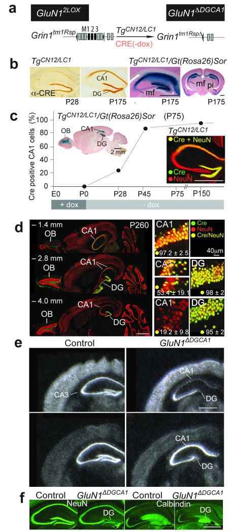
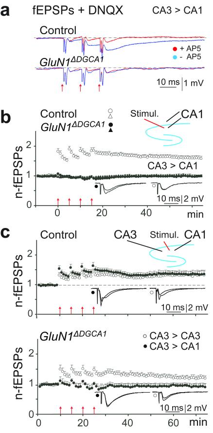
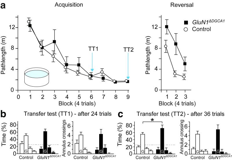
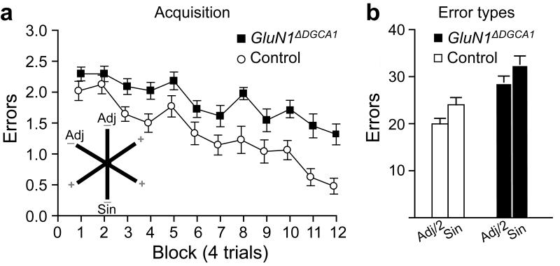
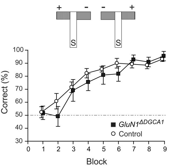
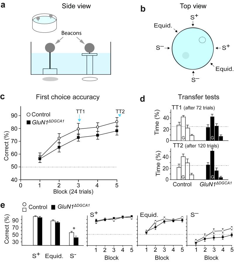
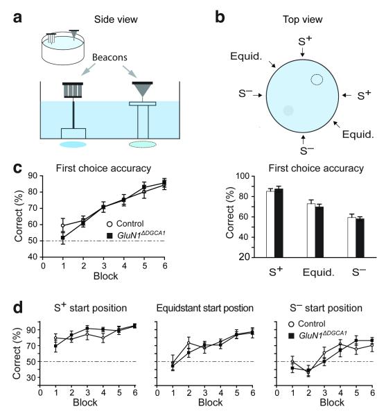
Hippocampal NMDA receptors (NMDARs) and NMDAR-dependent synaptic plasticity are widely considered crucial substrates of long-term spatial memory, although their precise role remains uncertain. Here we show that Grin1(ΔDGCA1) mice, lacking GluN1 and hence NMDARs in all dentate gyrus and dorsal CA1 principal cells, acquired the spatial reference memory water maze task as well as controls, despite impairments on the spatial reference memory radial maze task. When we ran a spatial discrimination water maze task using two visually identical beacons, Grin1(ΔDGCA1) mice were impaired at using spatial information to inhibit selecting the decoy beacon, despite knowing the platform's actual spatial location. This failure could suffice to impair radial maze performance despite spatial memory itself being normal. Thus, these hippocampal NMDARs are not essential for encoding or storing long-term, associative spatial memories. Instead, we demonstrate an important function of the hippocampus in using spatial knowledge to select between alternative responses that arise from competing or overlapping memories.


Figures

Figure 1
Figure 1. Removal of NMDARs in DG and CA1
(a) Genes to generate conditional NMDAR−/− mice (GluN1ΔDGCA1) by postnatal dox-controlled activation of TgCN12/LC1-encoded Cre in Grin1tm1Rsp mice (GluN12lox). LoxP sites are depicted by black triangles. Exons appear as boxes, and when black, encode membrane-spanning segments. (b) Anti-Cre immunostainings of brain sections from dox treated (till P0) TgCN12/LC1 mice at postnatal days P28 and P175, and β-galactosidase stainings (blue) of sections from TgCN12/LC1/Gt(Rosa)26Sor mice (mf, mossy fibers; pi, piriform cortex with ~30% of layer II neurons expressing Cre). (c, d) Dox-regulated Cre expression in the brains of TgCN12/LC1 mice. (c) Expression pattern of the nuclearly localized Cre in CA1 pyramidal cells when dox had been removed from the drinking water of mothers at the day of delivery. At P28 (n=2 mice), P45 (n=2) and P150 (n=6), the extent of Cre expressing pyramidal neurons in the dorsal CA1 layer was estimated by α-Cre immunostains (green), counterstained with α-NeuN (red). The inset shows Cre-activated β-galactosidase expression in CA1 and DG of the hippocampus and the olfactory bulb (OB) in a brain section from TgCN12/LC1/Gt(Rosa)26Sor mice. (d, left), Horizontal brain sections of TgCN12/LC1 mice immunostained with α-Cre (green) and α-NeuN (red) at three different levels from bregma (– 1.4, – 2.8, and – 4.0 mm) showed a yellow gradient of Cre/NeuN immunoreactivity in the dorsal to ventral str. pyramidale of CA1, but not in the DG. The percentage of Cre positive cells among >1,000 NeuN positive neurons was evaluated at various depths in dorsal and ventral hippocampus, in slices from four P260 GluN1ΔDGCA1 mice, and is listed as mean ± SD. (d, right) High magnification examples of α-Cre/α-NeuN co-labeled CA1 and DG neurons (yellow) from sections given on the left. The transition between dorsal and ventral hippocampus in the rostro-caudal axis had the highest variance of Cre expressing NeuN positive cells (middle insets). Other parts of the hippocampal formation feature negligible numbers of Cre-positive cells (<1% co-labeling). (e) In situ hybridization with GluN1 and GluA1 probes. Scale bars, 2mm. (f) α-NeuN and α-Calbindin immunostainings.
Figure 2
Figure 2. Loss of functional NMDA receptors at CA3-to-CA1 synapses in the dorsal CA1 region
(a) Absence of NMDA responses in field recordings in acute brain slices from 10 to 12 month old GluN1ΔDGCA1 mice: Blue traces show the average (five repetitions at 0.1 Hz) of fEPSPs elicited by three synaptic activations at 100 Hz in the presence of the AMPA receptor blocker DNQX (20μM) in control (upper panel) and GluN1ΔDGCA1 (lower panel) mice. Red traces are the corresponding responses following subsequent superfusion with AP5 (50μM) to block NMDA receptors. Mean representative traces (averaged across 5 stimulations both before and after AP5) from a single slice from both a control and a GluN1ΔDGCA1 mouse. Experiments were repeated in 3 control (n=12 slices) and 2 GluN1ΔDGCA1 mice (n=8 slices). (b) Absence of LTP: Normalized and pooled fEPSP slopes evoked at CA1 radiatum and oriens synapses in slices from both control (open symbols) and GluN1ΔDGCA1 mice (filled symbols). Forty to 45 min after the last tetanization of the afferent fibres in the stratum radiatum in slices from five Controls, the average slope of the field EPSP (open circles) was 1.60 ± 0.07 (mean ± SEM; n=18 slices) of the pre-tetanic value, whereas the untetanized oriens pathway (triangles) was unchanged (0.97 ± 0.04). In slices from four GluN1ΔDGCA1 mice LTP was completely abolished (0.99 ± 0.03, n=14 slices), and evoked responses were not significantly different from those in the untetanized pathway (1.03 ± 0.04; p = 0.35). (c) LTP at CA3-CA3 and CA3-CA1 synapses: normalized and pooled fEPSPs before and after LTP induction at CA3-CA3 (open circles) and CA3-CA1 (filled circles) radiatum synapses. In slices from three control mice (upper panel), LTP was well-developed 40 – 45 min post-tetanization, with similar magnitudes in the two regions (CA3; 1.33 ± 0.12, CA1; 1.37 ± 0.11; n=11 slices; p = 0.77). The same experimental paradigm performed on slices from three GluN1ΔDGCA1 mice (lower panel) showed preserved LTP in CA3 whereas LTP in CA1 failed to develop (CA3; 1.20 ± 0.07, CA1: 0.94 ± 0.19; n=8 slices; p = 0.01). All means ± s.e.m. Arrows at the abscissa indicate the time points of tetanic stimulation. Insets show means of six consecutive synaptic responses in the tetanized pathway before and 45 min post-tetanization.
Figure 3
Figure 3. Associative long-term spatial reference memory in the Morris watermaze
(a) Similar pathlengths for Controls (n=12) and GluN1ΔDGCA1 mice (n=12) during Acquisition of the fixed location, hidden escape platform watermaze task (Acquisition; geno – F<1, geno x block – F(8,176)=1.64; p>0.10; 4 trials/block). When the platform was moved to the opposite quadrant of the pool (Reversal), the pathlengths of GluN1ΔDGCA1 mice were significantly longer than Controls (geno – F(1,22)=13.41; p<0.005). All means ± s.e.m. (b, c) Long-term memory performance during probe trials (Transfer tests) after 6 and 9 training blocks. Percentage time spent (left panel) and numbers of annulus crossings (right panel) in each quadrant (left to right; adjacent left, goal (G), adjacent right, opposite) for Controls and GluN1ΔDGCA1 mice. In transfer test 2 (TT2), GluN1ΔDGCA1 mice searched longer in the training quadrant than Controls (t(22)=2.24; * p<0.05). Broken line: chance levels of performance. In addition, the numbers of annulus crossings in each quadrant are given.
Figure 4
Figure 4. Associative long-term spatial reference memory on the radial maze task
(a) Inset: Mice were trained to discriminate between three rewarded (+) and three un-baited (−) arms. Errors per trial for Controls (n=12) and GluN1ΔDGCA1 mice (n=11) across 12 blocks of testing (4 trials per block). GluN1ΔDGCA1 mice were impaired at discriminating between the baited and non-baited arms, making significantly more spatial reference memory errors (F (1,21) = 30.42; p<0.0001). (b) Error types during spatial reference memory acquisition. Total number of errors into the single, non-rewarded arm (Sin) and the total number of errors into the pair of adjacent (Adj/2), non-rewarded arms. The number of errors into the pair of adjacent, un-baited arms was divided by two to correct for the number of arms. All means ± s.e.m.
Figure 5
Figure 5. Preserved visual discrimination learning in GluN1ΔDGCA1 mice
In the visual discrimination task both Controls (n=13) and GluN1ΔDGCA1 mice (n=11) learned to associate floor/wall color (grey vs. black/white stripes) with a reward (+) independent of its spatial position in the arms of the maze. The top part of the figure depicts the T-maze with the start arm (S) and both the rewarded (+) and non-rewarded (−) goal arms, which can be arranged in either of two possible configurations. There were 9 blocks of training (10 trials per block). Mean ± s.e.m. percent correct choices for Controls and GluN1ΔDGCA1 mice. Broken line: chance levels of performance.
Figure 6
Figure 6. Spatial memory and choice performance on the spatial discrimination beacon watermaze task
(a) Schema and horizontal side view of the spatial beacon watermaze task. (b) Top view indicating the six possible start sites and the positions of the beacons (broken line = + Platform; shaded circle = – platform). (c) Both Controls (n=11) and GluN1ΔDGCA1 mice (n=12) learned to choose the correct beacon in terms of first choice accuracy: geno – F(1,21)=2.88; p>0.10, geno x blocks – F<1; 24 trials/block). (d) When probe tests (TT1 and TT2; beacons and platform removed) were conducted, both groups showed a strong preference for the goal quadrant (adjacent left, goal (G), adjacent right, opposite). (e) GluN1ΔDGCA1 mice were impaired at choosing the correct beacon when starting from a point close to the incorrect beacon (S), but not when starting from either of the other two start locations; (S+) or equidistant; geno x start position - F(2,42)=4.24; p<0.025, simple main effects group difference *p<0.025). (e, right) Mean ± s.e.m. percent correct choices across 5 blocks of training (8 trials/block) on trials starting from the S+, the equidistant and the S position. Broken line, chance levels of performance. All means ± s.e.m.
Figure 7
Figure 7. Normal performance of GluN1ΔDGCA1 mice on the visual discrimination beacon watermaze task
(a) Horizontal side view of the non-spatial beacon watermaze task. (b) Top view of the six possible start positions. (c) Both Controls (n=8) and GluN1ΔDGCA1 mice (n=9) learned to choose the correct beacon. First choice accuracy improved with training for both groups across 6 blocks of training (24 trials/block; c, left). The first choice accuracy across all training trials from different starting positions showed that all mice were less accurate at choosing the correct beacon when starting from S (c, right). (d) Performance across 6 blocks of training on trials starting from S+, equidistant and S positions. All mice performed less accurately when starting from S. There was no difference between Controls and GluN1ΔDGCA1 mice across 6 blocks of training for any of the start positions (8 trials/block). Broken line: chance levels of performance. All means ± s.e.m.

Comment in

References

    1. Bliss TV, Collingridge GL. A synaptic model of memory: long-term potentiation in the hippocampus. Nature. 1993;361:31–9. - PubMed
    1. Martin SJ, Grimwood PD, Morris RG. Synaptic plasticity and memory: an evaluation of the hypothesis. Annu. Rev. Neurosci. 2000;23:649–711. - PubMed
    1. Neves G, Cooke SF, Bliss TV. Synaptic plasticity, memory and the hippocampus: a neural network approach to causality. Nat. Rev. Neurosci. 2008;9:65–75. - PubMed
    1. Morris RG, Anderson E, Lynch GS, Baudry M. Selective impairment of learning and blockade of long-term potentiation by an N-methyl-D-aspartate receptor antagonist, AP5. Nature. 1986;319:774–776. - PubMed
    1. Bannerman DM, Good MA, Butcher SP, Ramsay M, Morris RG. Distinct components of spatial learning revealed by prior training and NMDA receptor blockade. Nature. 1995;378:182–186. - PubMed

Publication types