Review
. 2012 Oct;194(20):5495-503.
doi: 10.1128/JB.00304-12. Epub 2012 Jul 13.

Peroxide-sensing transcriptional regulators in bacteria

Affiliations
Review

Peroxide-sensing transcriptional regulators in bacteria

James M Dubbs et al. J Bacteriol. 2012 Oct.

Abstract

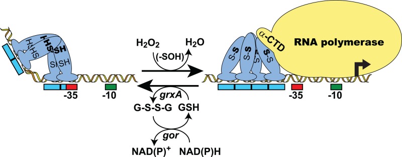
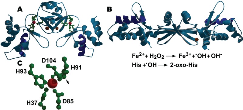
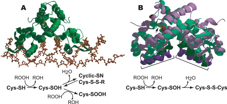
The ability to maintain intracellular concentrations of toxic reactive oxygen species (ROS) within safe limits is essential for all aerobic life forms. In bacteria, as well as other organisms, ROS are produced during the normal course of aerobic metabolism, necessitating the constitutive expression of ROS scavenging systems. However, bacteria can also experience transient high-level exposure to ROS derived either from external sources, such as the host defense response, or as a secondary effect of other seemingly unrelated environmental stresses. Consequently, transcriptional regulators have evolved to sense the levels of ROS and coordinate the appropriate oxidative stress response. Three well-studied examples of these are the peroxide responsive regulators OxyR, PerR, and OhrR. OxyR and PerR are sensors of primarily H(2)O(2), while OhrR senses organic peroxide (ROOH) and sodium hypochlorite (NaOCl). OxyR and OhrR sense oxidants by means of the reversible oxidation of specific cysteine residues. In contrast, PerR senses H(2)O(2) via the Fe-catalyzed oxidation of histidine residues. These transcription regulators also influence complex biological phenomena, such as biofilm formation, the evasion of host immune responses, and antibiotic resistance via the direct regulation of specific proteins.


Figures

Fig 1
Fig 1
Mechanism of transcription activation via intramolecular disulfide formation in 2-Cys OxyR. A model for H2O2-dependent, OxyR-mediated transcription activation of a target gene in E. coli is shown. Activation begins with the oxidation of the sensing cysteine (SH) residue of OxyR to sulfenic acid (-SOH), followed by the rapid formation of an intramolecular disulfide bond with the resolving cysteine (SH). The resulting conformational change often causes a shift in the DNase I footprint and can also affect DNA binding affinity and promoter conformation as well as render OxyR capable of interacting with RNA polymerase. Transcription activation involves the direct interaction of OxyR with the alpha subunit C-terminal domain (α-CTD) of RNA polymerase. Oxidized OxyR is reduced, using reduced glutathione (GSH) as the electron donor, via the glutaredoxin (grxA)/glutathione reductase (gor) system, with reducing equivalents ultimately supplied by NAD(P)H. Red and green boxes indicate RNA polymerase σ70 −35 and −10 promoter elements, respectively. Blue boxes indicate OxyR DNA binding contacts. Activation can also occur via oxidative modification of the sensing cysteine alone.
Fig 2
Fig 2
H2O2-mediated inactivation of PerR. (A) The ribbon structure of the carbon backbone of reduced B. subtilis PerR-Zn2+-Mn2+, showing the side chains of the amino acids coordinating the regulatory metal (green) and the DNA binding helices (dark blue), is shown. The structural and regulatory metals, Zn2+ and Mn2+, are shown as brown and red spheres, respectively. In its reduced state, PerR-Zn2+-Fe2+/Mn2+ binds to sites overlapping target promoter/operators and blocks transcription. (B) The ribbon structure of oxidized PerR-Zn2+-Mn2+ is shown. The steps in the Fenton-mediated oxidation of His residues in PerR-Zn2+-Fe2+ by H2O2 are shown below the panel. Exposure to H2O2 results in the iron-catalyzed production of OH, followed by the oxidation of the 2-carbon of the imidazole ring of one of two histidine residues (H37 and H91) that participate in coordinating the bound Fe2+ in each monomer. While H37 is the preferential target for oxidation, oxidation of either histidine to 2-oxo-histidine results in the disruption of normal Fe coordination, resulting in the destabilization of the DNA binding domain of the monomer and leading to the release of oxidized PerR from the DNA. (C) Close-up view of the Mn2+-containing regulatory metal binding site in reduced PerR that shows the relative positions of the metal-coordinating side groups of H37, H91, H93, D85, and D104 (positions of the coordinating side groups roughly correspond to those on the right side of panel A). The arrow indicates the 2-carbon of the imidazole group of H91, which is the site of oxidation in H91 and H37. In all cases, DNA binding helices are shown in dark blue. The Swiss Protein Data Bank (PDB) identifications (IDs) for reduced and oxidized PerR are 3F8N and 2RGV, respectively.
Fig 3
Fig 3
Organic hydroperoxide (ROOH)-mediated derepression of OhrR-regulated promoters. (A) The structure of reduced 1-Cys B. subtilis OhrR (green ribbon) bound to the ohrA operator (DNA sugar phosphate backbone in brown) is shown. The redox cycle of B. subtilis OhrR is depicted below. Reduced 1-Cys OhrR binds to the target promoter/operator through the interaction of winged helix-turn-helix DNA binding domains with the DNA major groove, thereby blocking transcription. In the presence of organic hydroperoxide (ROOH), the single sensing cysteine in reduced OhrR, C15 (yellow), is oxidized to cysteine sulfenic acid (Cys-SOH). The Cys-SOH derivative remains bound to the promoter and must undergo one of several further modifications to induce the conformational change necessary to release the repressor. Cys-SOH can react either with a reduced cellular thiol to form a mixed thiol (Cys-S-S-R) or with the amino group of a neighboring amino acid residue to form a cyclic amide (Cys-SN). Both Cys-S-S-R and Cys-SN are recycled in vivo via reduction. Derepression can also occur through further oxidation of the Cys-SOH, for example, to cysteine sulfinic acid (Cys-SOOH). Overoxidized derivatives of OhrR are likely degraded. (B) An overlay of the ribbon structures of the reduced (purple) and oxidized (green) forms of the 2-Cys X. campestris OhrR showing the peroxide-induced shift in the positions of the DNA binding helices (dark purple and dark green, indicated by brackets). The redox-active cysteines in reduced OhrR are colored brown, while the disulfide bonds in the oxidized form are rendered in yellow (also indicated by an arrow in the right-hand monomer). The redox cycle of a 2-Cys OhrR is depicted below. The sensing cysteine (C22) of reduced 2-Cys OhrR bound to a target promoter/operator is oxidized in the presence of ROOH to cysteine sulfenic acid (Cys-SOH). The sensing Cys-SOH rapidly reacts with a second “resolving” cysteine (C127) residue to form an intersubunit disulfide bond that induces a conformational change that repositions the DNA binding helices and releases OhrR from the promoter. The oxidized disulfide bond-containing form of 2-Cys OhrR is likely actively rereduced in vivo. The PDB IDs for reduced B. subtilis OhrR proteins bound to the ohrA operator and oxidized and reduced X. campestris OhrR proteins are 1Z9C, 2PFB, and 2PEX, respectively.

Similar articles

Cited by

References

    1. Antelmann H, Helmann JD. 2011. Thiol-based redox switches and gene regulation. Antioxid. Redox Signal. 14: 1049–1063 - PMC - PubMed
    1. Aslund F, Zheng M, Beckwith J, Storz G. 1999. Regulation of the OxyR transcription factor by hydrogen peroxide and the cellular thiol-disulfide status. Proc. Natl. Acad. Sci. U. S. A. 96: 6161–6165 - PMC - PubMed
    1. Atichartpongkul S, Fuangthong M, Vattanaviboon P, Mongkolsuk S. 2010. Analyses of the regulatory mechanism and physiological roles of Pseudomonas aeruginosa OhrR, a transcription regulator and a sensor of organic hydroperoxides. J. Bacteriol. 192: 2093–2101 - PMC - PubMed
    1. Bae HW, Cho YH. 2012. Mutational analysis of Pseudomonas aeruginosa OxyR to define the regions required for peroxide resistance and acute virulence. Res. Microbiol. 163: 55–63 - PubMed
    1. Ballal A, Ray B, Manna AC. 2009. sarZ, a sarA family gene, is transcriptionally activated by MgrA and is involved in the regulation of genes encoding exoproteins in Staphylococcus aureus. J. Bacteriol. 191: 1656–1665 - PMC - PubMed

Publication types

LinkOut - more resources