. 2012 Sep;194(18):5020-33.
doi: 10.1128/JB.00915-12. Epub 2012 Jul 13.

Transcriptional regulation of the ecp operon by EcpR, IHF, and H-NS in attaching and effacing Escherichia coli

Affiliations

Transcriptional regulation of the ecp operon by EcpR, IHF, and H-NS in attaching and effacing Escherichia coli

Verónica I Martínez-Santos et al. J Bacteriol. 2012 Sep.

Abstract

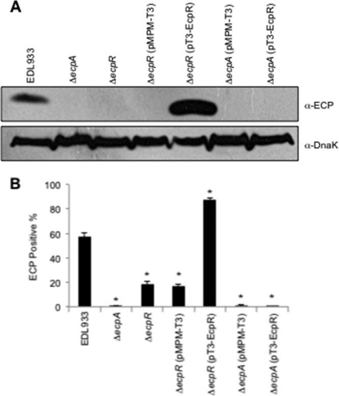
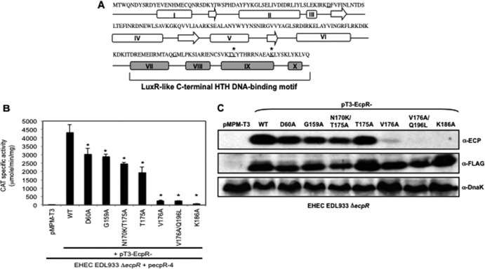
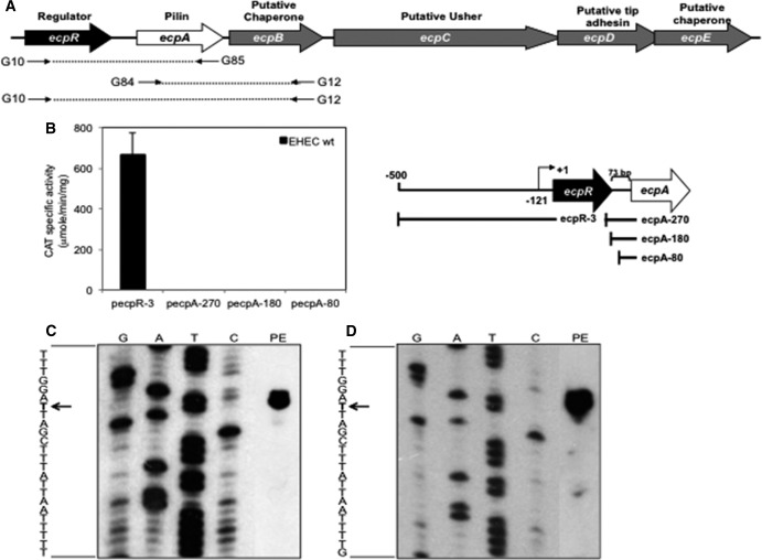
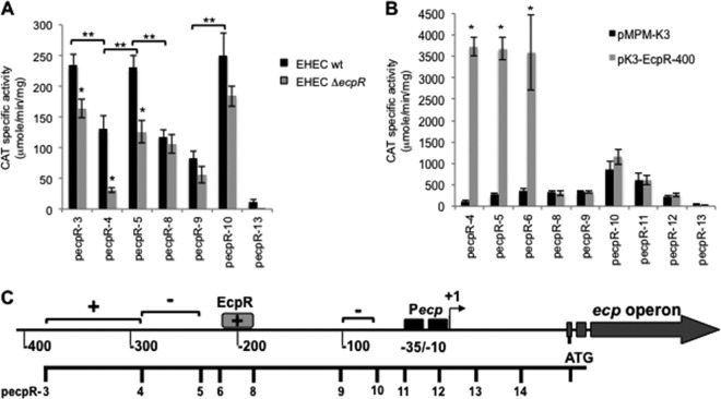
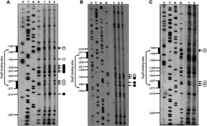
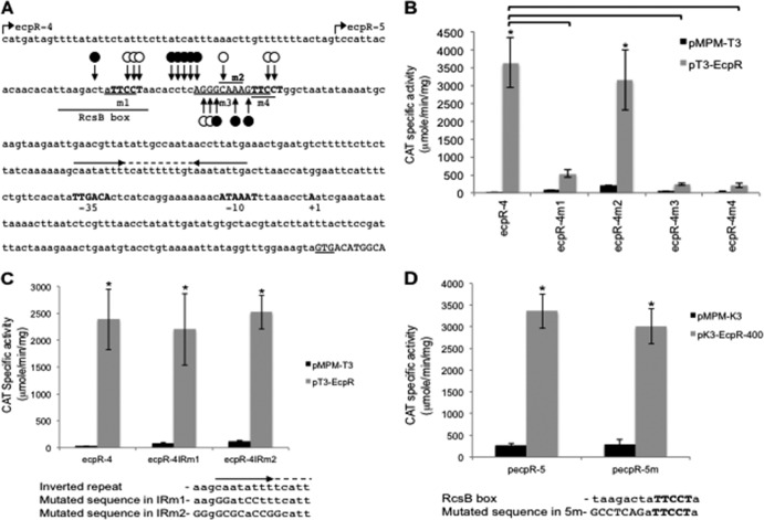
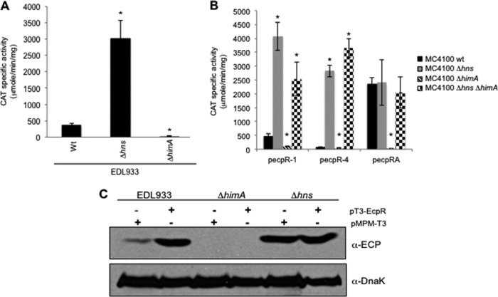
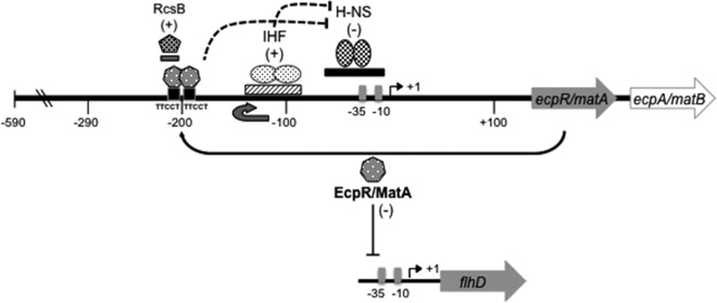
Enteropathogenic (EPEC) and enterohemorrhagic (EHEC) Escherichia coli are clinically important diarrheagenic pathogens that adhere to the intestinal epithelial surface. The E. coli common pili (ECP), or meningitis-associated and temperature-regulated (MAT) fimbriae, are ubiquitous among both commensal and pathogenic E. coli strains and play a role as colonization factors by promoting the interaction between bacteria and host epithelial cells and favoring interbacterial interactions in biofilm communities. The first gene of the ecp operon encodes EcpR (also known as MatA), a proposed regulatory protein containing a LuxR-like C-terminal helix-turn-helix (HTH) DNA-binding motif. In this work, we analyzed the transcriptional regulation of the ecp genes and the role of EcpR as a transcriptional regulator. EHEC and EPEC ecpR mutants produce less ECP, while plasmids expressing EcpR increase considerably the expression of EcpA and production of ECP. The ecp genes are transcribed as an operon from a promoter located 121 bp upstream of the start codon of ecpR. EcpR positively regulates this promoter by binding to two TTCCT boxes distantly located upstream of the ecp promoter, thus enhancing expression of downstream ecp genes, leading to ECP production. EcpR mutants in the putative HTH DNA-binding domain are no longer able to activate ecp expression or bind to the TTCCT boxes. EcpR-mediated activation is aided by integration host factor (IHF), which is essential for counteracting the repression exerted by histone-like nucleoid-structuring protein (H-NS) on the ecp promoter. This work demonstrates evidence about the interplay between a novel member of a diverse family of regulatory proteins and global regulators in the regulation of a fimbrial operon.


Figures

Fig 1
Fig 1
EcpR is required for the synthesis of ECP. (A) Western blot analysis of whole-cell extracts of EHEC EDL933 wild type, ΔecpA, ΔecpR, ΔecpR/pMPM-T3, ΔecpR/pT3-EcpR, ΔecpA/pMPM-T3, and ΔecpA/pT3-EcpR (Table 1) grown in DMEM at 30°C in static cultures. DnaK was detected as a loading control. (B) Production of ECP by wild-type EHEC and its isogenic ecpA and ecpR mutants, analyzed by flow cytometry using anti-ECP antibodies and goat anti-rabbit IgG Alexa Fluor 488 conjugate; 10,000 events were measured. Bacteria were recovered from the supernatant of HeLa cells infected for 6 h at 37°C. Error bars represent the standard deviations of three independent assays done in duplicate. *, P < 0.02 between wild-type and mutant strains, calculated using the unpaired Student t test.
Fig 2
Fig 2
Mutations in the predicted HTH DNA-binding domain of EcpR affect its function. (A) Schematic representation of the predicted secondary structure of EcpR using the PSIPRED server. The arrows indicate β strands, and the rectangles represent α helices. Amino acids replaced by alanine are underlined. The asterisks indicate residues that affect EcpR activity when replaced by alanines. Helices VII, VIII, IX, and X corresponding to the putative EcpR HTH DNA-binding domain are shown in gray. (B) CAT activity assay with EDL933 ΔecpR containing fusion ecpR-4 complemented with vector pMPM-T3 or plasmids encoding wild-type EcpR (pT3-EcpR) or the EcpR mutants (pT3-EcpR-D60A, pT3-EcpR-G159A, pT3-EcpR-N170KT175A, pT3-EcpR-T175A, pT3-EcpR-V176A, pT3-EcpR-V176AQ196L, and pT3-EcpR-K186A). The resulting strains were grown in DMEM at 37°C with shaking for 6 h. Error bars represent the standard deviations of the activity of three independent assays done in duplicate. *, P < 0.0001 between mutants and wild-type EcpR. (C) Western blot of whole-cell extracts of EHEC ΔecpR transformed with vector pMPM-T3 or with plasmids encoding wild-type EcpR or the mutants. EcpR was detected with α-FLAG antibodies. DnaK was detected as a loading control.
Fig 3
Fig 3
The ecpRABCDE genes are transcribed as an operon. (A) Schematic representation of the ecp cluster formed by genes ecpR and ecpA to ecpE. The putative function of each gene is indicated above the big arrows. The arrows below the schematic represent the primers used for RT-PCR, and the dashed lines represent the products obtained with each pair of primers. (B) CAT activity produced by EHEC EDL933 transformed with fusions ecpR-3, ecpA-270, ecpA-180, and ecpA-80 (left panel). Error bars indicate standard deviations of results from three independent experiments with duplicates. The right panel is the schematic representation of the ecpR and ecpA-cat transcriptional fusions used in the left panel. The broken arrow indicates the transcriptional start site. The ecpA gene is separated from ecpR by 73 bp. As a negative control, the strain transformed with vector pKK232-8 was used. (C and D) Identification of the transcriptional start site. Total RNA from the wild-type EHEC strain EDL933 (C) and EPEC E2348/69 (D) transformed with fusions ecpRA-H and ecpRA-P, respectively, was extracted from DMEM culture samples collected at an OD600 of 1.0. A primer specific for the 5′ end of the coding region of ecpR (positions +136 to +156, with respect to the transcriptional start site) and 10 μg of total RNA were used for the primer extension reaction, as indicated in Materials and Methods. The arrows indicate the bases corresponding to the transcriptional start sites.
Fig 4
Fig 4
Identification of regulatory elements involved in ecp regulation. (A) Expression of cat transcriptional fusions contained in plasmids ecpR-3, ecpR-4, ecpR-5, ecpR-8, ecpR-9, ecpR-10, and ecpR-13 in wild-type EHEC EDL933 and its ΔecpR isogenic mutant. (B) Expression of cat transcriptional fusions contained in plasmids ecpR-4, ecpR-5, ecpR-6, ecpR-8, ecpR-9, ecpR-10, ecpR-11, ecpR-12, and ecpR-13 in EHEC ΔecpR also carrying vector pMPM-K3 or plasmid pK3-EcpR-400. CAT-specific activity was determined from samples obtained from static DMEM cultures grown at 30°C. Error bars represent the standard deviations of the activity of three independent assays done in duplicate. *, P < 0.0001 between gray and black bars in panels A and B. **, P < 0.0001 between black bars in panel A. (C) Schematic representation of the regulatory region of ecp. The broken arrow indicates the transcriptional start site, and the black boxes represent the −10 and −35 boxes. Brackets represent positive (+) and negative (−) regulatory elements. The gray box spans the proposed EcpR-binding site according to the deletion analysis. The scale below the schematic indicates the 5′ end of each fusion.
Fig 5
Fig 5
Identification of the EcpR-binding site. (A to C) In vivo DMS footprinting analysis of the EHEC ecp regulatory region. Bacterial cultures of EHEC EDL933 ΔecpR containing fusion ecpR-4 (A and B) or ecpR-4m2 (C) plus either the empty vector pMPM-T3 (lanes 1) or plasmids expressing wild-type EcpR (pT3-EcpR, lanes 2) or the EcpR K186A inactive mutant (pT3-EcpR-K186A, lanes 3) were exposed to DMS, and the plasmid DNA was extracted and treated as described in Materials and Methods. Primer extension products were amplified with primers pKK-8-BHI (A and C) and ecpR-3R (B) and resolved by 7% polyacrylamide–8 M urea gel electrophoresis and visualized by autoradiography. White circles and black circles indicate protected and hypermethylated sites, respectively. Black vertical bars on the left side indicate the bases spanning the putative EcpR-binding sites. The sequencing ladder was obtained with the same primers.
Fig 6
Fig 6
Analysis of ecp regulatory elements by site-directed mutagenesis. (A) Sequence of the regulatory region of ecp. The EcpR start codon (GTG) is underlined. The transcriptional start site, the −10 and −35 hexamers, and the putative binding sites for EcpR, corresponding to the TTCCT boxes, are indicated in bold uppercase letters. The bases changed in mutants ecpR-4m1 to ecpR-4m4 are underlined, as well as the RcsB box. The broken arrows represent the 5′ limit of the ecpR-4 and ecpR-5 fusions at positions −288 and −236, respectively. Black circles represent the hypermethylated bases observed by in vivo footprinting, while white circles represent protected bases. The horizontal arrows indicate the locations of an inverted repeat, and the dashed line indicates the 11-bp spacer. (B) Analysis of the TTCCT boxes. CAT activity of fusions ecpR-4, ecpR-4m1, ecpR-4m2, ecpR-4m3, and ecpR-4m4. Changes in each mutant fusion are indicated in panel A. (C) Analysis of the inverted repeats. CAT activity of fusions ecpR-4, ecpR-4IRm1 and ecpR4IRm2. The wild-type and mutant sequences of the region spanning the left arm of the inverted repeat are shown below. (D) Analysis of the RcsB box. CAT activity of fusions pecpR-5 and pecpR-5m. The wild-type and mutant sequences of the RcsB box are shown below. The bases changed in fusion pecpR-5m are indicated with capital letters. CAT activity was determined from culture samples of EHEC EDL933 ΔecpR complemented with the empty vector pMPM-T3 (B and C) or pMPM-K3 (D) or plasmid pT3-EcpR (B and C) or pK3-EcpR-400 (D) plus the different transcriptional fusions. The activity is the result of three independent experiments done in duplicate. Asterisks (*) correspond to P values of <0.0001 between gray and black bars. Brackets correspond to P values of <0.0001 between gray bars.
Fig 7
Fig 7
Global regulators IHF and H-NS regulate ecp expression. (A) CAT activities of fusion ecpR-1 in wild-type EHEC EDL933 and its Δhns and ΔhimA isogenic mutants. *, P values of <0.0001. (B) CAT activity assay with E. coli K-12 strain MC4100 and its Δhns, ΔhimA, and ΔhimA Δhns isogenic mutants transformed with fusions ecpR-1, ecpR-4, and ecpRA. Error bars indicate standard deviations of results from three independent experiments with duplicates. *, P values of <0.0001 between wild-type and mutant strains, calculated using the unpaired Student t test. (C) Western blot with the anti-ECP antibody of whole-cell extracts of EHEC EDL933 and its Δhns and ΔhimA isogenic mutants transformed with vector pMPM-T3 or plasmid pT3-EcpR (Table 1). DnaK was detected as a loading control using a monoclonal anti-DnaK antibody.
Fig 8
Fig 8
Transcriptional regulation of the ecp (mat) operon. ecp (mat) codes for EcpR/MatA, a regulatory protein containing a LuxR_C-like DNA-binding HTH domain that regulates positively the expression of the ecp operon by binding to two TTCCT boxes distantly located at positions −211 to −207 (distal box) and −189 to −185 (proximal box) with respect to the TSS. EcpR and IHF, which most likely bends the DNA between the TTCCT boxes and the promoter to get EcpR into close proximity to the promoter, counteract the repression exerted by H-NS on the ecp (mat) promoter. In addition, the response regulator RcsB also positively regulates the ecp (mat) promoter by binding to a sequence that overlaps the distant TTCCT box (40). EcpR/MatA also acts as a negative regulator by repressing the expression of the flagellar master operon flhDC (39).

Similar articles

Cited by

References

    1. Agron PG, Ditta GS, Helinski DR. 1993. Oxygen regulation of nifA transcription in vitro. Proc. Natl. Acad. Sci. U. S. A. 90:3506–3510 - PMC - PubMed
    1. Aravind L, Anantharaman V, Balaji S, Babu MM, Iyer LM. 2005. The many faces of the helix-turn-helix domain: transcription regulation and beyond. FEMS Microbiol. Rev. 29:231–262 - PubMed
    1. Avelino F, et al. 2010. The majority of enteroaggregative Escherichia coli strains produce the E. coli common pilus when adhering to cultured epithelial cells. Int. J. Med. Microbiol. 300:440–448 - PubMed
    1. Baga M, Goransson M, Normark S, Uhlin BE. 1985. Transcriptional activation of a pap pilus virulence operon from uropathogenic Escherichia coli. EMBO J. 4:3887–3893 - PMC - PubMed
    1. Barba J, et al. 2005. A positive regulatory loop controls expression of the locus of enterocyte effacement-encoded regulators Ler and GrlA. J. Bacteriol. 187:7918–7930 - PMC - PubMed

Publication types

MeSH terms

LinkOut - more resources