. 2013 Mar;65(2):157-72.
doi: 10.1007/s10616-012-9481-y. Epub 2012 Jul 14.

Beyond annexin V: fluorescence response of cellular membranes to apoptosis

Affiliations

Beyond annexin V: fluorescence response of cellular membranes to apoptosis

Alexander P Demchenko. Cytotechnology. 2013 Mar.

Abstract

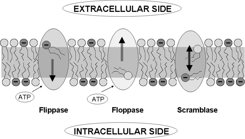
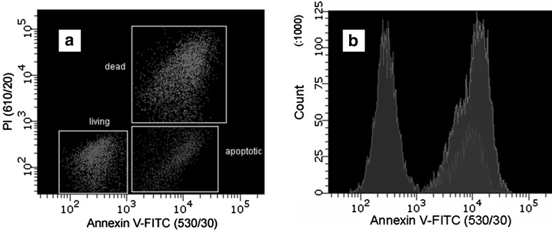
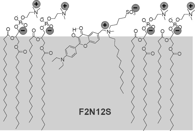
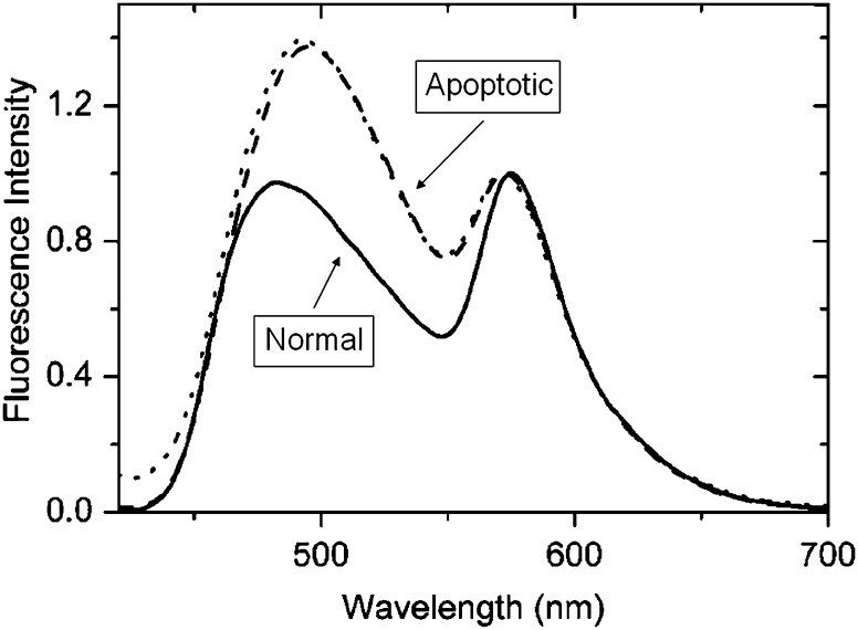
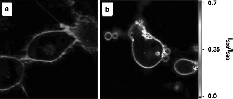
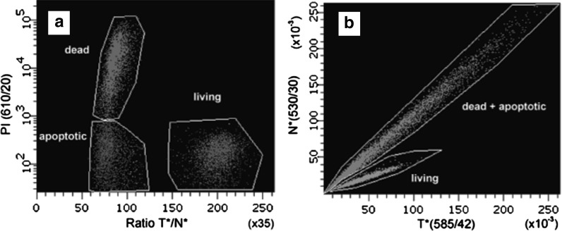
Dramatic changes in the structure of cell membranes on apoptosis allow easy, sensitive and non-destructive analysis of this process with the application of fluorescence methods. The strong plasma membrane asymmetry is present in living cells, and its loss on apoptosis is commonly detected with the probes interacting strongly and specifically with phosphatidylserine (PS). This phospholipid becomes exposed to the cell surface, and the application of annexin V labeled with fluorescent dye is presently the most popular tool for its detection. Several methods have been suggested recently that offer important advantages over annexin V assay with the ability to study apoptosis by spectroscopy of cell suspensions, flow cytometry and confocal or two-photon microscopy. The PS exposure marks the integrated changes in the outer leaflet of cell membrane that involve electrostatic potential and hydration, and the attempts are being made to provide direct probing of these changes. This review describes the basic mechanisms underlying the loss of membrane asymmetry during apoptosis and discusses, in comparison with the annexin V-binding assay, the novel fluorescence techniques of detecting apoptosis on cellular membrane level. In more detail we describe the detection method based on smart fluorescent dye F2N12S incorporated into outer leaflet of cell membrane and reporting on apoptotic cell transformation by easily detectable change of the spectral distribution of fluorescent emission. It can be adapted to any assay format.


Figures

Fig. 1
Fig. 1
Transporter-controlled exchange of phospholipids between intracellular and extracellular leaflets of the cell membrane creating strong charge asymmetry (Zwaal et al. 2005), modified
Fig. 2
Fig. 2
Comparison of “labeling” and “sensing” methodologies in application of fluorescence dyes
Fig. 3
Fig. 3
The principle of apoptosis detection method based on annexin V. With the aid of Ca2+ ions, this protein interacts with high affinity with PS heads exposed on the membrane surface. Annexin V can be labeled with fluorescent dye that allows visualization of cells exposing PS
Fig. 4
Fig. 4
Flow cytometry of T lymphoblastoid cells treated by actinomycin D and stained by annexin V-FITC and PI. FITC and PI were selectively excited at 488 nm and their fluorescence at 530 and 610 nm, respectively, was collected simultaneously. a Cells are sorted into living (dark green), dead (purple), and apoptotic (blue) cell populations by the absolute intensities of the two probes. Note that on both coordinates the scale is logarithmic. b Histograms plotted on the basis of intensity response of bound annexin V-FITC (Shynkar et al. 2007). (Color figure online)
Fig. 5
Fig. 5
The principle of apoptosis detection method based on F2N12S dye. Due to high affinity, the dye is incorporated into the phospholipid moiety of the outer leaflet of the membrane and reports on the changes in surface potential, molecular order and hydration by the change of color of its emission. On apoptosis the relative intensity of “green” emission increases over the “orange” emission. (Color figure online)
Fig. 6
Fig. 6
Location of F2N12S probe in the outer leaflet of biological membrane. Interactions between opposite charges with PC molecules are indicated
Fig. 7
Fig. 7
Fluorescence spectra of F2N12S probe in cell suspensions. The spectra were recorded in normal (solid) or in apoptotic CEM cells either in the absence (dashes) or in the presence of 2 mM Ca2+ ions (dots). Excitation wavelength was 400 nm. Final probe concentration was 100 nM (Shynkar et al. 2007)
Fig. 8
Fig. 8
Example of an application of F2N12S in cell microscopy. Fluorescence ratiometric images of normal (a) and apoptotic (b) cells stained with F2N12S using two-photon excitation at 830 nm. Note that the images are presented in artificial color based on intensity ratio at 520 and 580 nm (Oncul et al. 2010). (Color figure online)
Fig. 9
Fig. 9
Example of an application of F2N12S in flow cytometry. a The distribution of treated cells stained by F2N12S between the living (green) and apoptotic plus dead cells can be seen as two separate populations based on T*/N* ratio. Application of logarithmic coordinate based on PI response allows separating apoptotic (blue) and dead (purple) cell sub-populations. b Representation of results of the same experiment in two coordinates of N* and T* intensities for living (green) and dead plus apoptotic (red) cells. The wavelengths of maximal transmission and bandwidths of used filters are indicated in parenthesis for the headings of the axes (Shynkar et al. 2007). (Color figure online)

Similar articles

Cited by

References

    1. Alam IS, Neves AA, Witney TH, Boren J, Brindle KM. Comparison of the C2A domain of synaptotagmin-I and annexin-V as probes for detecting cell death. Bioconjug Chem. 2010;21:884–891. doi: 10.1021/bc9004415. - DOI - PubMed
    1. Altschuh D, Oncul S, Demchenko AP. Fluorescence sensing of intermolecular interactions and development of direct molecular biosensors. J Mol Recognit. 2006;19:459–477. doi: 10.1002/jmr.807. - DOI - PubMed
    1. Balasubramanian K, Schroit AJ. Aminophospholipid asymmetry: a matter of life and death. Annu Rev Physiol. 2003;65:701–734. doi: 10.1146/annurev.physiol.65.092101.142459. - DOI - PubMed
    1. Balasubramanian K, Bevers EM, Willems GM, Schroit AJ. Binding of annexin V to membrane products of lipid peroxidation. Biochemistry. 2001;40:8672–8676. doi: 10.1021/bi010841y. - DOI - PubMed
    1. Blankenberg FG, Norfray JF. Multimodality molecular imaging of apoptosis in oncology. Am J Roentgenol. 2011;197:308–317. doi: 10.2214/AJR.11.6953. - DOI - PubMed

LinkOut - more resources