. 2012 Oct;26(8):644-55.
doi: 10.1007/s12149-012-0631-2. Epub 2012 Jul 15.

F-18 fluorodeoxyglucose uptake and water-perfusable tissue fraction in assessment of myocardial viability

Affiliations

F-18 fluorodeoxyglucose uptake and water-perfusable tissue fraction in assessment of myocardial viability

Hidehiro Iida et al. Ann Nucl Med. 2012 Oct.

Abstract

Objectives: (15)O-water-perfusable tissue fraction (PTF) has been shown to be a potential index for assessing myocardial viability in PET, an alternative to (18)F-fluorodeoxyglucose (FDG). This study aimed to directly compare these two independent methods in assessing myocardial viability in patients with abnormal wall motion.

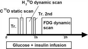
Methods: PET study was performed on 16 patients with previous myocardial infarction, before coronary artery bypass graft operation (CABG). The protocol included a (15)O-carbonmonoxide static, a (15)O-water dynamic and an (18)F-FDG dynamic scan, during the euglycemic hyperinsulinemic clamp. Echocardiography was performed at the time of PET and 5-12 months after the CABG, and the wall motion recovery was evaluated on segmental and global bases. Consistency between PTF and (18)F-FDG was evaluated visually and also in a quantitative manner. Predictive values for the wall motion recovery were also compared between the two approaches.

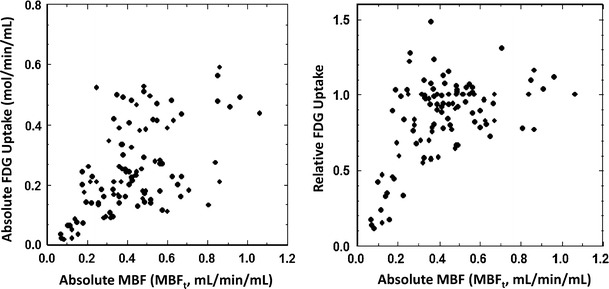
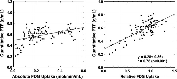
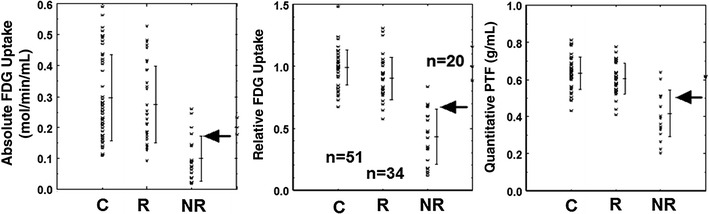
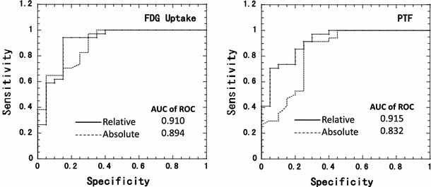
Results: The image quality of (18)F-FDG was superior to that of (15)O-water. The qualitative PTF showed significantly smaller defects than (18)F-FDG, and the quantitative PTF showed slightly greater values than (18)F-FDG in the infarcted region. The two methods were, however, consistent visually and also quantitatively. The predictive values of the wall motion recovery were almost equal between the two approaches. The absolute (18)F-FDG uptake was varied in normal segments, and predictive values for the wall motion recovery by the absolute (18)F-FDG was less (accuracy: 80 %) compared with those by the relative (18)F-FDG (accuracy: 87 %) and the quantitative PTF (accuracy: 89 %).

Conclusion: Despite the small sample size, PTF appears to give consistent results with the (18)F-FDG approach, and might be an alternative viability assessment.


Figures

Fig. 1
Fig. 1
Schematic diagram of the study protocol. Tr indicates the transmission scan. The second transmission scan was used to confirm that the patient did not move during the period
Fig. 2
Fig. 2
Example images of extra-vascular tissue density (D ev), the blood volume (V B), 15O-water washout phase (WO, or qualitative PTF), and 18F-FDG uptake, obtained from 2 typical cases. In case 1, the wall motion was irreversible in the anterior wall segment (arrow, a), which was well predicted by both WO and FDG as complete defect. In case 2, wall motion was reduced in the anterior wall (arrow, b), but was not improved in the apex (arrow, c). These were also consistent with the findings in both WO and FDG. Image quality of 18F-FDG was better than that of WO, but both images provided consistent results
Fig. 3
Fig. 3
Relation between 18F-FDG uptake and absolute myocardial blood flow determined by means of 15O-water PET. Left: absolute uptake of 18F-FDG which was calculated using the arterial input function. Right: relative uptake of 18F-FDG to a control region. MBFt denotes ml of blood per minutes per ml of regions-of-interest adopted to this plot
Fig. 4
Fig. 4
Relation between the water-perfusable tissue fraction (PTF, g/ml) versus 18F-FDG uptake (Left: absolute 18F-FDG uptake. Right: relative 18F-FDG uptake)
Fig. 5
Fig. 5
Absolute and relative 18F-FDG uptake and water-perfusable tissue fraction (PTF) in control segments (C) and in revascularized segments. Arrows correspond to the threshold values to discriminate between the recovered (R) and non-recovered (NR) (see text)
Fig. 6
Fig. 6
ROC analysis for wall motion recovery after successful coronary artery bypass graft surgery in patients with hypocontractile function

References

    1. Rahimtoola SH. A perspective on the three large multicenter randomized clinical trials of coronary bypass surgery for chronic stable angina. Circulation. 1985;72(6 Pt 2):V123–V135. - PubMed
    1. Braunwald E, Rutherford JD. Reversible ischemic left ventricular dysfunction: evidence for the “hibernating myocardium”. J Am Coll Cardiol. 1986;8(6):1467–1470. doi: 10.1016/S0735-1097(86)80325-4. - DOI - PubMed
    1. Rahimtoola SH. The hibernating myocardium. Am Heart J. 1989;117(1):211–221. doi: 10.1016/0002-8703(89)90685-6. - DOI - PubMed
    1. Marshall RC, Tillisch JH, Phelps ME, et al. Identification and differentiation of resting myocardial ischemia and infarction in man with positron computed tomography, 18F-labeled fluorodeoxyglucose and N-13 ammonia. Circulation. 1983;67(4):766–778. doi: 10.1161/01.CIR.67.4.766. - DOI - PubMed
    1. Schwaiger M, Brunken R, Grover-McKay M, et al. Regional myocardial metabolism in patients with acute myocardial infarction assessed by positron emission tomography. J Am Coll Cardiol. 1986;8(4):800–808. doi: 10.1016/S0735-1097(86)80420-X. - DOI - PubMed

Publication types

MeSH terms

LinkOut - more resources