. 2012 Jul 31;109(31):12592-7.
doi: 10.1073/pnas.1204710109. Epub 2012 Jul 13.

Neuroepithelial body microenvironment is a niche for a distinct subset of Clara-like precursors in the developing airways

Affiliations

Neuroepithelial body microenvironment is a niche for a distinct subset of Clara-like precursors in the developing airways

Arjun Guha et al. Proc Natl Acad Sci U S A. .

Abstract

Clara cells of mammalian airways have multiple functions and are morphologically heterogeneous. Although Notch signaling is essential for the development of these cells, it is unclear how Notch influences Clara cell specification and if diversity is established among Clara cell precursors. Here we identify expression of the secretoglobin Scgb3a2 and Notch activation as early events in a program of secretory cell fate determination in developing murine airways. We show that Scgb3a2 expression in vivo is Notch-dependent at early stages and ectopically induced by constitutive Notch1 activation, and also that in vitro Notch signaling together with the pan-airway transcription factor Ttf1 (Nkx2.1) synergistically regulate secretoglobin gene transcription. Furthermore, we identified a subpopulation of secretory precursors juxtaposed to presumptive neuroepithelial bodies (NEBs), distinguished by their strong Scgb3a2 and uroplakin 3a (Upk3a) signals and reduced Ccsp (Scgb1a1) expression. Genetic ablation of Ascl1 prevented NEB formation and selectively interfered with the formation of this subpopulation of cells. Lineage labeling of Upk3a-expressing cells during development showed that these cells remain largely uncommitted during embryonic development and contribute to Clara and ciliated cells in the adult lung. Together, our findings suggest a role for Notch in the induction of a Clara cell-specific program of gene expression, and reveals that the NEB microenvironment in the developing airways is a niche for a distinct subset of Clara-like precursors.


Conflict of interest statement

The authors declare no conflict of interest.

Figures

Fig. 1.
Fig. 1.
Spatial pattern of Notch activation in the developing airways. (A–G) Labeling for cleaved Notch1 (NICD, red) and Ascl1 (green) indicates that Notch signaling is activated in the airways a few days earlier than Ccsp expression is detected (E15.5, 14), and that it is active in the NEB microenvironment from early stages. (A and B) Low levels of NICD were detected at E12.5 in trachea (Tr) and main bronchi (arrow), but not in distal (dl) airways (shown at higher resolution in B). (C–F) At E14.5, NICD labeling was more abundant in both the extrapulmonary airways (Tr in C, higher magnification in D) and proximal intrapulmonary airways (C, higher magnification in E), but no labeling was detected in distal airways (C Inset). (F) Clusters of strong NICD labeling in the proximal airways at E14.5 are associated with clusters of Ascl1-expressing cells (arrows, airway outlined in white). Note the solitary Ascl1-expressing cells (arrowhead) are not associated with strong NICD labeling (F). (G) At E18.5, widespread labeling of NICD was detected in the intrapulmonary airways, including the NEB microenvironment (arrow). L, lumen; Lu, lung; V, vasculature. (Scale bar: 10 μm.)
Fig. 2.
Fig. 2.
Scgb3a2 expression correlates with NICD and identifies a distinct subpopulation of Clara-like progenitors associated with pNEBs. (A–D) Time course of Scgb3a2 expression revealed a pattern similar to that of NICD (Fig. 1). Scgb3a2 was first detected in the trachea (Tr) at E12.5 (A) and in the intrapulmonary airways from E13.5–E14.5 (B–D). At E18.5 (D), Scgb3a2 expression was widespread and detected in both trachea and terminal bronchiole (TB). Clusters of cells with strong signal could be discerned at E13.5–E14.5 (B and C Inset, arrows). (E) Colocalization of Scgb3a2 and NICD was readily observed in these cell clusters (arrows; L marks the airway lumen). (F) Double-Scgb3a2 ISH/AsclI IHC revealed Scgb3a2-labeled clusters (F Right) juxtaposed to Ascl1-expressing pNEBs. (F Right Inset) Scgb3a2-expressing cells (blue) have a clear nucleus not labeled by anti-Ascl1 (brown). No Ascl1-expressing cells were detected in the trachea (F Left). (G) High-resolution optical section showing that luminal Scgb3a2-expressing cells (ISH, red) could be distinguished from basal Ascl1-expressing cells (IHC, green). (H) Scgb3a2 and Ascl1 double-labeling at E13.5 suggests that the formation of Ascl1 clusters (distal, arrows) precedes formation of Scgb3a2-Ascl1 dual clusters. (I–O) Analysis of Ccsp and Scgb3a2 expression at E18.5 suggested that the cells associated with pNEBs may be a distinct subpopulation of Clara precursors. (I–K) Labeling of NICD (red, shown separately in J), Ccsp (white, shown separately in K), and Ascl1 (green) showed that the cells in the NEB microenvironment are NICD positive, but express low (arrow)-to-negligible levels of Ccsp at this stage. Cells with both NICD and Ccsp were abundant away from the NEB microenvironment (I). (L–O) Triple labeling for Scgb3a2 (ISH, red, shown separately in M), Ccsp (ISH, green, shown separately in N), and Cgrp (IHC, blue, shown separately in O) at E18.5 showed that cells apposed to pNEBs express Scgb3a2 and low Ccsp (arrowhead), and some have Scgb3a2 but negligible Ccsp (arrow). Elsewhere, Scgb3a2 and Ccsp signals are strong and colocalized (L).
Fig. 3.
Fig. 3.
High levels of Upk3a expression distinguishes Clara-like precursors in the pNEB microenvironment. (A) Profiling of E18.5 control and Notch signaling-deficient (Rbpjcnull) lungs identified known markers for Clara cells and implicated Upk3a as a candidate marker. (B and C) ISH at E18.5 in control lungs revealed that Upk3a was highly enriched in clusters of cells in the proximal airways (C, arrow) and expressed at low levels in scattered cells in distal airways (C, bracket). No signal was detected along the airway axis in Rbpjcnull airways (Tr, trachea). (D) qPCR analysis showed that Upk3a levels can be detected at E12.5 and increase throughout development. (E and F) Upk3a transcripts were detected by ISH from E14.5 onward in cell clusters juxtaposed to Ascl1-expressing cells (E and F; Insets show higher magnification). Some expression away from clusters was detected from E16.5 onward (F, bracket). Upk3a expression was also detected in a few cells in the adult airways (G, arrow) frequently juxtaposed to Cgrp-expressing NEBs (G Inset). (H–K) Upk3a expression was perturbed in Ascl1-null (Ascl1−/−) lungs. Clusters of high Upk3a expression were detected in control at both E14.5 (H) and E18.5 (J), but not in mutant lungs at either time point [I and K, circled regions; note expression of Upk3A is detected in the esophagus in the mutant at E14.5 (I, eso)]. Rare Upk3a-expressing cells (nonclustered) could still be seen in the E18.5 Ascl1-null mutant (K Inset). (L) qPCR analysis of Upk3a expression in Rbpjk-deficient and Ascl1 mutants at E14.5 and E18.5 showing that Upk3a expression is dependent on both Notch signaling and Ascl1.
Fig. 4.
Fig. 4.
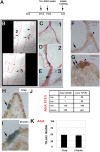
Lineage analysis of Upk3a-expressing cells at E15.5 reveals that these cells are precursors of Clara and ciliated cells. (A) Experimental protocol for the induction and harvest of tissue from the Upk3acreERT2 × Rosa26lacz. (B–E) Upk3a lineage-derived cells (blue) are distributed along the proximal/distal axis (counterstained with Fast Red) in clusters and as solitary cells (B, arrows). Regions 1, 2, and 3 in B are shown at higher magnification in C–E. (F and G) Double labeling of X-gal–stained preparations for PGP9.5 shows that Upk3a lineage-derived cells (arrows) are not in close association with NEBs (arrowheads), although rare examples of apposition are observed (G). Note that LacZ (arrow)- and PGP9.5 (arrowhead)- expressing cells are distinct. (H and I) Upk3a-expressing cells labeled at E15.5 contribute to Clara and ciliated lineages in adults. Double labeling of X-gal–stained preparations for Ccsp (H) and β-tubulin (I) demonstrate that these cells contribute to both Clara (H) and ciliated (I) lineages. (J and K) Quantitation of the numbers of X-gal–stained cells that co-label for Clara and ciliated markers at E18.5 (Ccsp, Foxj1, n = 156 airways) and in adults (Ccsp, β-Tubulin, n = 501 airways). The Upk3a lineage-derived cells are mostly uncommitted to either Clara or ciliated fates at E18.5 but differentiate into these lineages thereafter. K, mean ± SEM.
Fig. 5.
Fig. 5.
Notch-dependent regulation of secretory cell-associated genes. (A–F) Analysis of Notch gain and loss of function in E14.5 airways in vivo. (A and B) Disruption of Notch signaling in Rbpjcnull mutants disrupts expression of Scgb3a2 and Upk3a. Comparable segments of the intrapulmonary airways in A and B (encircled by gray lines) evidence negligible expression of both markers in Rbpjcnull lungs. Residual low-level expression of Scgb3a2 was detected in the extrapulmonary airways of mutants only after prolonged staining (A, arrow). Double-Scgb3a2 ISH/AsclI IHC shows that specification of pNEBs was unaffected in Rbpjcnull mutants, but no Scgb3a2 was detected in surrounding cells (A Inset, arrowhead). (C–F) Shh cre-driven NICD perturbed lung architecture (compare distribution of Ttf1 in C and D) and resulted in widespread expression of Scgb3a2 and Upk3a. (G and H) In vitro analysis of the role of Notch signaling in the regulation of the Scgb3a2 promoter. (G) Schematic showing presumptive Rbpjk binding sites (high-affinity site, red arrow; low-affinity site, gray arrow) in the Scgb3a2 promoter 1 kb upstream to the transcription start site. (G Lower) Sequence of a −273-bp fragment of the Scgb3a2 promoter showing putative Rbpjk (red) and TTF1 (green) binding sites. (H) Validation of the predicted high-affinity Rbpjk binding site in the Scgb3a2 promoter by EMSA. An anti-Rbpjk antibody supershifted the labeled oligonucleotide containing the putative high-affinity binding site (CTRL, lane +ab). Note that the band that is supershifted by anti-Rbpjk was competed by coincubation of the labeled oligonucleotide with an unlabeled CTRL oligonucleotide (cognate) but not competed by coincubation with an unlabeled mutant oligonucleotide lacking the high-affinity binding site (MUT, sequences shown below). (I) Luciferase assay examining the sufficiency of NICD and Ttf1 in the transactivation of the Scgb3a2 promoter. NICD expression alone did not significantly transactivate the Scgb3a2 promoter, but coexpression of NICD and Ttf1 synergistically up-regulated reporter expression. This synergistic up-regulation was abolished when the high-affinity Rbpjk binding site was mutated (−273mut; G).

References

    1. Rawlins EL, et al. Epithelial stem/progenitor cells in lung postnatal growth, maintenance, and repair. Cold Spring Harb Symp Quant Biol. 2008;73:291–295. - PubMed
    1. Rawlins EL, et al. The role of Scgb1a1+ Clara cells in the long-term maintenance and repair of lung airway, but not alveolar, epithelium. Cell Stem Cell. 2009;4:525–534. - PMC - PubMed
    1. Reynolds SD, Malkinson AM. Clara cell: Progenitor for the bronchiolar epithelium. Int J Biochem Cell Biol. 2010;42:1–4. - PMC - PubMed
    1. Stripp BR, Maxson K, Mera R, Singh G. Plasticity of airway cell proliferation and gene expression after acute naphthalene injury. Am J Physiol. 1995;269:L791–L799. - PubMed
    1. Hung KS. Development of neuroepithelial bodies in pre- and postnatal mouse lungs: Scanning electron microscopic study. Anat Rec. 1982;203:285–291. - PubMed

Publication types

MeSH terms