Review
. 2012 Jul 1;125(Pt 13):3075-83.
doi: 10.1242/jcs.092353. Epub 2012 Jul 13.

Molecular force transduction by ion channels: diversity and unifying principles

Affiliations
Review

Molecular force transduction by ion channels: diversity and unifying principles

Sergei Sukharev et al. J Cell Sci. .

Abstract

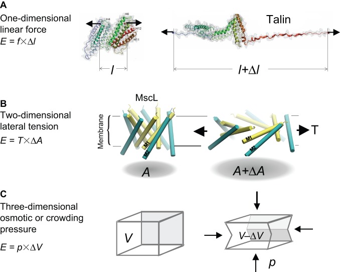
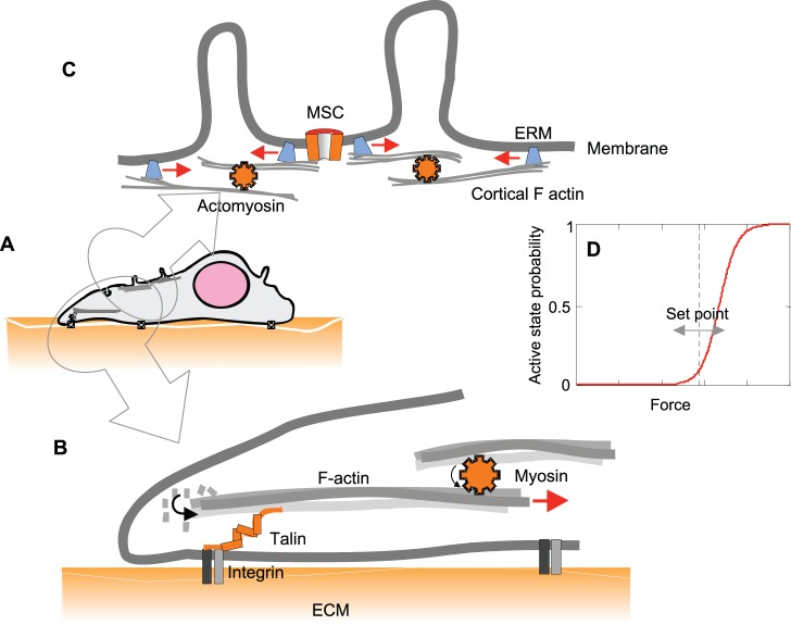
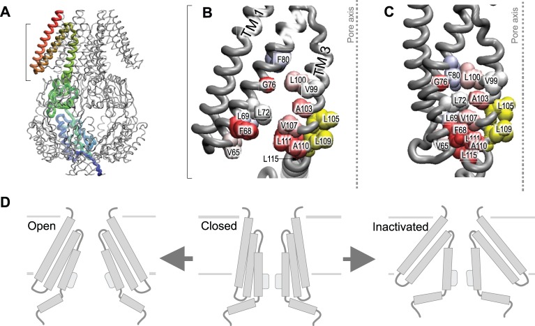
Cells perceive force through a variety of molecular sensors, of which the mechanosensitive ion channels are the most efficient and act the fastest. These channels apparently evolved to prevent osmotic lysis of the cell as a result of metabolite accumulation and/or external changes in osmolarity. From this simple beginning, nature developed specific mechanosensitive enzymes that allow us to hear, maintain balance, feel touch and regulate many systemic variables, such as blood pressure. For a channel to be mechanosensitive it needs to respond to mechanical stresses by changing its shape between the closed and open states. In that way, forces within the lipid bilayer or within a protein link can do work on the channel and stabilize its state. Ion channels have the highest turnover rates of all enzymes, and they can act as both sensors and effectors, providing the necessary fluxes to relieve osmotic pressure, shift the membrane potential or initiate chemical signaling. In this Commentary, we focus on the common mechanisms by which mechanical forces and the local environment can regulate membrane protein structure, and more specifically, mechanosensitive ion channels.


Figures

Fig. 1.
Fig. 1.
Mechanosensing in different dimensions. (A) A one-dimensional sensor, such as talin, can unfold and elongate with tension. Such a conformational change could expose cryptic binding sites within the protein [modified with permission from del Rio et al., 2009. Reprinted with permission from AAAS. (del Rio et al., 2009)]. (B) A membrane channel that opens in response to membrane tension is an example of a two-dimensional sensor that increases its in-plane area (A). The panel shows the predicted tilting motion of pairs of transmembrane helices of the bacterial mechanosensitive channel MscL, which are associated with a ∼20 nm2 change of in-plane area expansion [modified with permission from Sukharev et al., 2005, ASM Press, Washington, DC (Sukharev et al., 2005)]. (C) A hypothetical elastic structure that decreases its volume under osmotic pressure is an example of a three-dimensional mechanosensor.
Fig. 2.
Fig. 2.
Pre-stressing sensors affects their sensitivity through mechanical adaptation. (A) An adherent cell spreads lamellipodia that probe the stiffness of the substrate by applying force to focal adhesions. (B) Diagrammatic representation of the actin–talin–integrin linkage, in which partial unfolding of talin that was pre-stressed through actomyosin leads to reinforcement of focal adhesions (reviewed by Moore et al., 2010). (C) Blebbing of the plasma membrane mediated by actomyosin, which is connected to the membrane through anchoring to ERM family proteins, at the same time, might pre-stress adjacent membranes that contain a mechanosensitive channel (MSC). The diagram is based on the ideas discussed by Thery and Bornens (Thery and Bornens, 2008). (D) Dose–response curve of an MSC. The curve has a sigmoid shape (Boltzmann distribution), in which the slope sensitivity (the change in active state probability for a given change in force) depends on the resting force. This defines the set-point and the dynamic range of response.
Fig. 3.
Fig. 3.
Adaptive gating mechanism of the bacterial small-conductance MSC (MscS). (A) The crystal structure of the MscS (PDB ID 2OAU) heptamer with one subunit highlighted in color. The TM2–TM3 helices show an open crevice (B) and a closed crevice that has been generated by computational rearrangement (C). The TM2–TM3 hydrophobic link might serve as a ‘clutch’ that connects the peripheral helices to the gate (yellow). The Lys111Ser mutant shows a strong propensity toward inactivation as a result of compromised TM2–TM3 interactions, which transmit force from the membrane to the gate. Figures reproduced with permission from (Akitake et al., 2005; Belyy et al., 2010a).

Similar articles

Cited by

References

    1. Akinlaja J., Sachs F. (1998). The breakdown of cell membranes by electrical and mechanical stress. Biophys. J. 75, 247–254 10.1016/S0006-3495(98)77511-3 - DOI - PMC - PubMed
    1. Akitake B., Anishkin A., Sukharev S. (2005). The “dashpot” mechanism of stretch-dependent gating in MscS. J. Gen. Physiol. 125, 143–154 10.1085/jgp.200409198 - DOI - PMC - PubMed
    1. Alford P. W., Dabiri B. E., Goss J. A., Hemphill M. A., Brigham M. D., Parker K. K. (2011). Blast-induced phenotypic switching in cerebral vasospasm. Proc. Natl. Acad. Sci. USA 108, 12705–12710 10.1073/pnas.1105860108 - DOI - PMC - PubMed
    1. Anishkin A., Sukharev S. (2004). Water dynamics and dewetting transitions in the small mechanosensitive channel MscS. Biophys. J. 86, 2883–2895 10.1016/S0006-3495(04)74340-4 - DOI - PMC - PubMed
    1. Anishkin A., Akitake B., Kamaraju K., Chiang C. S., Sukharev S. (2010). Hydration properties of mechanosensitive channel pores define the energetics of gating. J. Phys. Condens. Matter 22, 454120 10.1088/0953-8984/22/45/454120 - DOI - PubMed

Publication types