Review
. 2012 Jul 1;125(Pt 13):3015-24.
doi: 10.1242/jcs.079509. Epub 2012 Jul 13.

Deconstructing the third dimension: how 3D culture microenvironments alter cellular cues

Affiliations
Review

Deconstructing the third dimension: how 3D culture microenvironments alter cellular cues

Brendon M Baker et al. J Cell Sci. .

Abstract

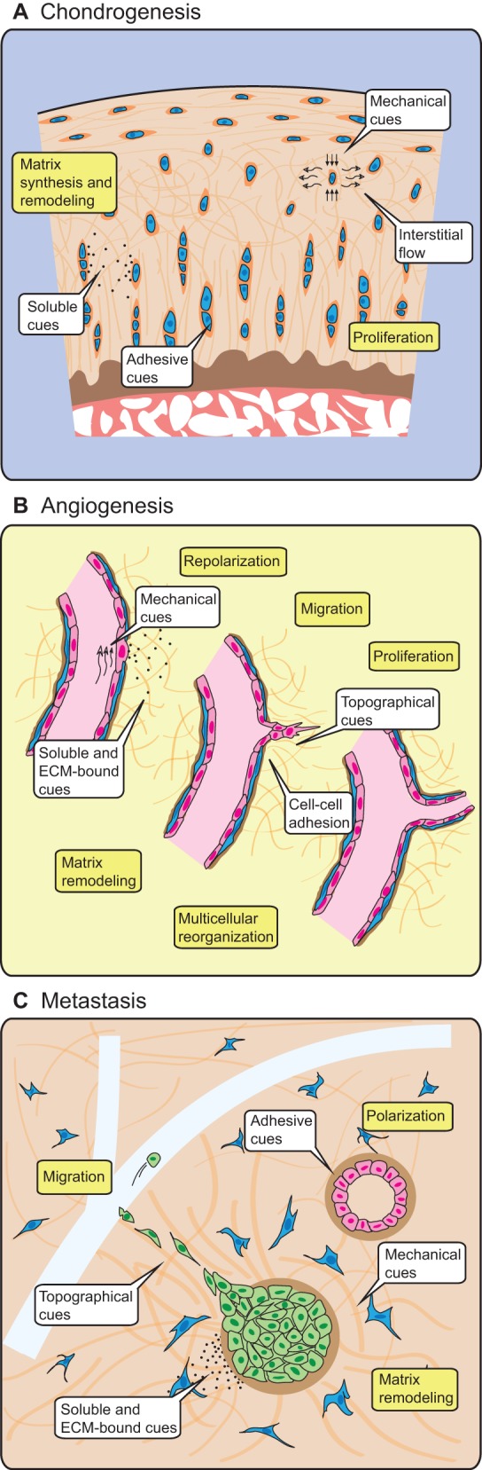
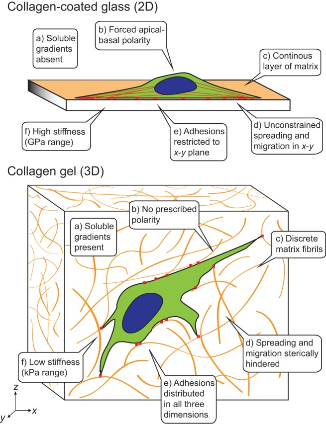
Much of our understanding of the biological mechanisms that underlie cellular functions, such as migration, differentiation and force-sensing has been garnered from studying cells cultured on two-dimensional (2D) glass or plastic surfaces. However, more recently the cell biology field has come to appreciate the dissimilarity between these flat surfaces and the topographically complex, three-dimensional (3D) extracellular environments in which cells routinely operate in vivo. This has spurred substantial efforts towards the development of in vitro 3D biomimetic environments and has encouraged much cross-disciplinary work among biologists, material scientists and tissue engineers. As we move towards more-physiological culture systems for studying fundamental cellular processes, it is crucial to define exactly which factors are operative in 3D microenvironments. Thus, the focus of this Commentary will be on identifying and describing the fundamental features of 3D cell culture systems that influence cell structure, adhesion, mechanotransduction and signaling in response to soluble factors, which - in turn - regulate overall cellular function in ways that depart dramatically from traditional 2D culture formats. Additionally, we will describe experimental scenarios in which 3D culture is particularly relevant, highlight recent advances in materials engineering for studying cell biology, and discuss examples where studying cells in a 3D context provided insights that would not have been observed in traditional 2D systems.


Figures

Fig. 1.
Fig. 1.
3D cellular phenomena in development, tissue homeostasis and disease are conducted by adhesive, mechanical and chemical cues originating from other cells and the extracellular environment. (A) Chondrocytes (blue) reside within a specialized pericellular ECM, where they are exposed to compressive forces, interstitial fluid flow, adhesive cues and soluble cues in the form of cytokines, which allow the cells to form and maintain the surrounding cartilage. (B) In response to soluble and matrix-bound growth factors and flow-induced mechanical forces on the blood vessel wall, endothelial cells (pink) alter their polarity and cell-cell contacts, and degrade the surrounding basement membrane (brown) and stromal ECM (orange) in order to collectively invade the surrounding tissue and form tubular sprouts. (C) The formation of normal epithelial structures (pink) requires adhesive and mechanical cues from neighboring cells and the basement membrane (brown) in order to tightly regulate proliferation and apoptosis. Misregulation of proliferation through genetic or extracellular changes initiates a cascade of soluble signals that activate fibroblasts (blue) in the surrounding stroma. Subsequent mechanical and structural changes in the stromal ECM enable transformed epithelial cells (green) to migrate towards neighboring vasculature (light blue) and, eventually, to metastasize. Drawings not to scale.
Fig. 2.
Fig. 2.
Adhesive, topographical, mechanical, and soluble cues in 2D and 3D. The cues encountered by a cell are strikingly different between an ECM-coated glass or plastic surface (2D) and a typical 3D ECM, such as collagen.

Similar articles

Cited by

References

    1. Adams R. H., Alitalo K. (2007). Molecular regulation of angiogenesis and lymphangiogenesis. Nat. Rev. Mol. Cell Biol. 8, 464–478 10.1038/nrm2183 - DOI - PubMed
    1. Amatangelo M. D., Bassi D. E., Klein–Szanto A. J. P., Cukierman E. (2005). Stroma-derived three-dimensional matrices are necessary and sufficient to promote desmoplastic differentiation of normal fibroblasts. Am. J. Pathol. 167, 475–488 10.1016/S0002-9440(10)62991-4 - DOI - PMC - PubMed
    1. Anselme K., Davidson P., Popa A. M., Giazzon M., Liley M., Ploux L. (2010). The interaction of cells and bacteria with surfaces structured at the nanometre scale. Acta Biomater. 6, 3824–3846 10.1016/j.actbio.2010.04.001 - DOI - PubMed
    1. Arnold M., Cavalcanti–Adam E. A., Glass R., Blümmel J., Eck W., Kantlehner M., Kessler H., Spatz J. P. (2004). Activation of integrin function by nanopatterned adhesive interfaces. ChemPhysChem 5, 383–388 10.1002/cphc.200301014 - DOI - PubMed
    1. Bashkin P., Doctrow S., Klagsbrun M., Svahn C. M., Folkman J., Vlodavsky I. (1989). Basic fibroblast growth factor binds to subendothelial extracellular matrix and is released by heparitinase and heparin-like molecules. Biochemistry 28, 1737–1743 10.1021/bi00430a047 - DOI - PubMed

Publication types

Substances