Review
. 2012 Jul 1;125(Pt 13):3061-73.
doi: 10.1242/jcs.093005. Epub 2012 Jul 13.

Mechanosensitive mechanisms in transcriptional regulation

Affiliations
Review

Mechanosensitive mechanisms in transcriptional regulation

Akiko Mammoto et al. J Cell Sci. .

Abstract

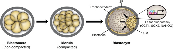
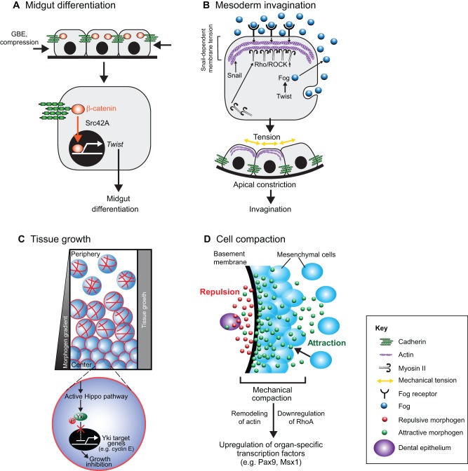
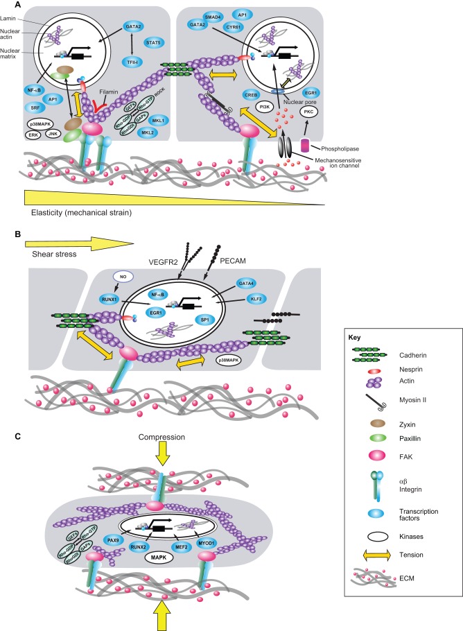
Transcriptional regulation contributes to the maintenance of pluripotency, self-renewal and differentiation in embryonic cells and in stem cells. Therefore, control of gene expression at the level of transcription is crucial for embryonic development, as well as for organogenesis, functional adaptation, and regeneration in adult tissues and organs. In the past, most work has focused on how transcriptional regulation results from the complex interplay between chemical cues, adhesion signals, transcription factors and their co-regulators during development. However, chemical signaling alone is not sufficient to explain how three-dimensional (3D) tissues and organs are constructed and maintained through the spatiotemporal control of transcriptional activities. Accumulated evidence indicates that mechanical cues, which include physical forces (e.g. tension, compression or shear stress), alterations in extracellular matrix (ECM) mechanics and changes in cell shape, are transmitted to the nucleus directly or indirectly to orchestrate transcriptional activities that are crucial for embryogenesis and organogenesis. In this Commentary, we review how the mechanical control of gene transcription contributes to the maintenance of pluripotency, determination of cell fate, pattern formation and organogenesis, as well as how it is involved in the control of cell and tissue function throughout embryogenesis and adult life. A deeper understanding of these mechanosensitive transcriptional control mechanisms should lead to new approaches to tissue engineering and regenerative medicine.


Figures

Fig. 1.
Fig. 1.
Mechanical transcriptional control and pluripotency. The expanding blastocoel pushes the inner cell mass (ICM) against the outer zona pellucida (ZP), which induces specific transcription factors (TFs) that are crucial for pluripotency.
Fig. 2.
Fig. 2.
Mechanical control of transcription during embryonic pattern formation. (A) Midgut differentiation. In Drosophila anterior stomodeal cells, mechanical compression as a result of germ band extension (GBE) induces expression of the gene encoding the transcription factor Twist, which is crucial for midgut differentiation through a Src42A-dependent mechanotransduction process that leads to the translocation of β-catenin into the nucleus. Modified with permission from Mammoto and Ingber, 2010 (Mammoto and Ingber, 2010). (B) Mesoderm invagination. In the Drosophila mesoderm, the Snail-dependent unstable pulses of apical constriction lead to a mechanically induced inhibition of Fog endocytosis through an increase in membrane tension. This activates the Fog–Rho–ROCK–Myo II signaling pathway, which leads to stable apical constriction and invagination. Fog is expressed under the control of Twist. Modified with permission from Mammoto and Ingber, 2010 (Mammoto and Ingber, 2010). (C) Tissue growth. During wing growth in Drosophila, cells in the central regions are compressed. This results in remodeling of actin in these cells, which activates the Hippo pathway and leads to phosphorylation of the transcription factor Yki, which prevents its translocation into the nucleus. Consequently, transcriptional activity of Yki target genes, such as cyclin E, is decreased and cell growth is inhibited. At the same time, cells at the periphery slow their proliferation because they have grown beyond the edges of the morphogen gradient. By employing these two mechanisms, the whole tissue grows uniformly. (D) Cell compaction. During teeth development in mouse, the early dental epithelium (DE) produces the antagonistic morphogens FGF8 (green) and semaphorin 3f (red) that attract and repulse mesenchymal cells, respectively. This causes mesenchymal cells to pack tightly during mesenchymal condensation in tooth development. The resultant mechanical compaction-induced changes in cell shape induce sets of transcription factors that are crucial for organ-specific morphogenesis.
Fig. 3.
Fig. 3.
Mechanotransduction pathways and transcriptional control in response to mechanical strain, fluid shear stress and compression. Cells and tissues are stabilized through a tensegrity mechanism in which a stabilizing tensional prestress or state of isometric tension is generated inside the cells by mechanical forces. The latter are generated by interactions of actin and myosin within the cytoskeleton that are worked against by integrin adhering to the ECM, cadherin adhering to neighboring cells and by internal cytoskeletal structures (Ingber, 2006). This mechanical equilibrium governs cell mechanics and hence, modulates the response of cells to external forces. (A) When cells and tissues experience mechanical strain, forces transmitted through integrin receptors and cadherins alter intracellular signaling pathways involving proteins such as kinases (e.g. PKC, MAPK14, ERKs and JNK), focal adhesion proteins (e.g. FAK, zyxin, paxillin) and Rho small GTPases and its guanine nucleotide exchange factors (GEFs) and GTPase activating proteins (GAPs). Mechanical tension exerted on integrins also modulates mechanosensitive ion channels and phospholipases, which activate PI3K and PKC, respectively, through second messengers such as intracellular Ca2+, inositol lipids and arachidonic acid. All of these mechanochemical signals lead to the activation of transcription factors (e.g. GATA2, TFII-I, NF-κB, AP1, SRF, STAT5, MKL1, MKL2, SMAD4, CYR61, CREB and EGR1). Physical forces that are exerted on surface adhesion receptors and are transmitted directly to the nucleus along cytoskeletal filaments and molecules that connect the cytoskeleton to the nucleus, such as nesprin, can influence transcription activity more directly. (B) When adherent cells, such as endothelial cells, are exposed to apical fluid shear stress, the cells sense these forces through mechanosensors such as VEGFR2 or PECAM. Various transcriptional activities (e.g. RUNX1, GATA4, KLF2, NF-κB, EGR1 and SP1) are modulated through downstream changes in biochemical signaling (e.g. NO, p38MAPK) and cytoskeletal prestress. (C) Compressive forces generated by gravity or physical movements that are transmitted across the ECM can also alter activities of intracellular biochemical signaling molecules (e.g. Rho GTPases, their GEFs and GAPs and MAPK) and cytoskeletal tension. Again, these changes can modulate various transcriptional activities (involving for example PAX9, RUNX2, MEF2 and MYOD1) that are crucial for homeostasis of tissues that normally bear compressive forces, such as bone, cartilage and tooth. Parts of this figure were modified with permission from Mammoto and Ingber, 2010 (Mammoto and Ingber, 2010).

Similar articles

Cited by

References

    1. Aarnes G. T., Steen H., Ludvigsen P., Kristiansen L. P., Reikerås O. (2002). High frequency distraction improves tissue adaptation during leg lengthening in humans. J. Orthop. Res. 20, 789–792 10.1016/S0736-0266(01)00175-9 - DOI - PubMed
    1. Adamo L., Naveiras O., Wenzel P. L., McKinney–Freeman S., Mack P. J., Gracia–Sancho J., Suchy–Dicey A., Yoshimoto M., Lensch M. W., Yoder M. C.et al. (2009). Biomechanical forces promote embryonic haematopoiesis. Nature 459, 1131–1135 10.1038/nature08073 - DOI - PMC - PubMed
    1. Agarwal S., Deschner J., Long P., Verma A., Hofman C., Evans C. H., Piesco N. (2004). Role of NF-kappaB transcription factors in antiinflammatory and proinflammatory actions of mechanical signals. Arthritis Rheum. 50, 3541–3548 10.1002/art.20601 - DOI - PMC - PubMed
    1. Ahmad M., Theofanidis P., Medford R. M. (1998). Role of activating protein-1 in the regulation of the vascular cell adhesion molecule-1 gene expression by tumor necrosis factor-alpha. J. Biol. Chem. 273, 4616–4621 10.1074/jbc.273.8.4616 - DOI - PubMed
    1. Alenghat F. J., Tytell J. D., Thodeti C. K., Derrien A., Ingber D. E. (2009). Mechanical control of cAMP signaling through integrins is mediated by the heterotrimeric Galphas protein. J. Cell. Biochem. 106, 529–538 10.1002/jcb.22001 - DOI - PMC - PubMed

Publication types

Substances