. 2012 Jul;245(7):369-80.
doi: 10.1007/s00232-012-9459-x. Epub 2012 Jul 15.

Extracellular loop cysteine mutant of cx37 fails to suppress proliferation of rat insulinoma cells

Affiliations

Extracellular loop cysteine mutant of cx37 fails to suppress proliferation of rat insulinoma cells

Miranda E Good et al. J Membr Biol. 2012 Jul.

Abstract

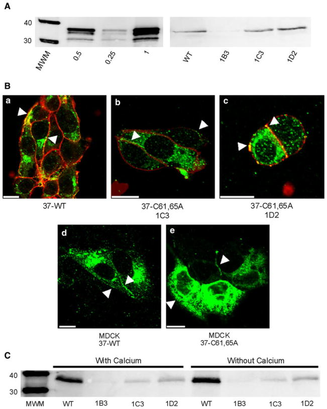
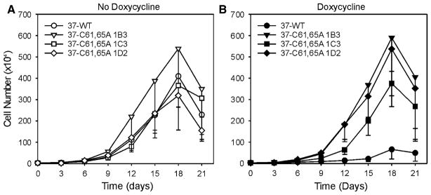
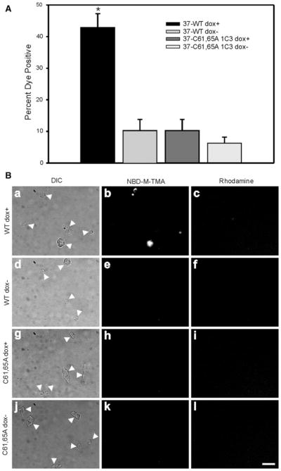
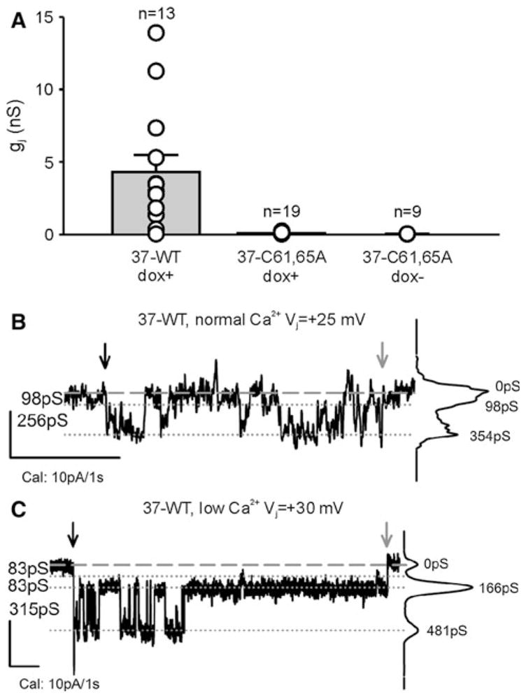
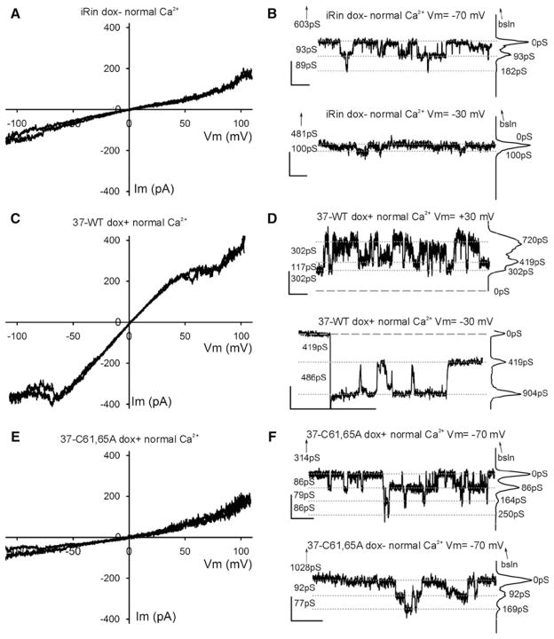
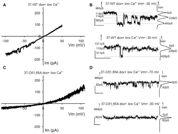
Although a functional pore domain is required for connexin 37 (Cx37)-mediated suppression of rat insulinoma (Rin) cell proliferation, it is unknown whether functional hemichannels would be sufficient or if Cx37 gap junction channels are required for growth suppression. To test this possibility, we targeted extracellular loop cysteines for mutation, expecting that the mutated protein would retain hemichannel, but not gap junction channel, functionality. Cysteines at positions 61 and 65 in the first extracellular loop of Cx37 were mutated to alanine and the mutant protein (Cx37-C61,65A) expressed in Rin cells. Although the resulting iRin37-C61,65A cells expressed the mutant protein comparably to Cx37 wild-type (Cx37-WT)--expressing Rin cells (iRin37), Cx37-C61,65A expression did not suppress the proliferation of Rin cells. As expected, iRin37-C61,65A cells did not form functional gap junction channels. However, functional hemichannels also could not be detected in iRin37-C61,65A cells by either dye uptake or electrophysiological approaches. Thus, failure of Cx37-C61,65A to suppress the proliferation of Rin cells is consistent with previous data demonstrating the importance of channel functionality to Cx37's growth-suppressive function. Moreover, failure of the Cx37-C61,65A hemichannel to function, even in low external calcium, emphasizes the importance of extracellular loop cysteines not only in hemichannel docking but also in determining the ability of the hemichannel to adopt a closed configuration that can open in response to triggers, such as low external calcium, effective at opening Cx37-WT hemichannels.


Figures

Fig. 1
Fig. 1
Expression and localization of Cx37-C61,65A and Cx37-WT are comparable. A Western blots of GST-37CT loaded in different amounts (left) and total protein isolated from iRin37 and three clones (1B3, 1C3 and 1D2) of iRin37-C61,65A cells (right). iRin cells were induced for 24 h with 2 μg/ml doxycycline, whole-cell protein was isolated and 50 μg of total cell protein was loaded for each sample. Expression levels for each cell line, determined by comparing band intensities of the samples to the standard curve generated using GST-37CT, were 3.7, 4.5 and 5.1 pmol/mg of total cell protein for clones 1B3, 1C3 and 1D2, respectively, and 4.5 pmol/mg protein for iRin37 cells. B Confocal images of Cx37 (green) localization with biotinylated membrane proteins (red) in induced iRin37 (37-WT) (a), iRin37-C61,65A 1C3 (37-C61,65A 1C3) (b) and iRin37-C61,65A 1D2 (37-C61,65A 1D2) (c) cells showing the presence of punctate labeling at appositional and nonappositional membranes (arrows). Confocal images of Cx37 (green) localization in MDCK cells transiently transfected to express Cx37-WT (MDCK 37-WT) (d) or Cx37-C61,65A (MDCK 37-C61,65A) (e) also showing the presence of punctate labeling at appositional and nonappositional membranes (arrows). Scale bars = 10 μm. C Western blot of membrane localized Cx37 isolated from Cx37-WT or -C61,65A expressing iRin cells. Induced iRin37 (WT) and iRin37-C61,65A clones 1B3, 1C3 and 1D2 were DTT- and maleimide-biotin-treated and harvested, and protein was immunoprecipitated with streptavidin and immunoblotted with anti-Cx37 antibody. Solutions either contained or lacked calcium. Detected Cx37 indicates membrane insertion of this protein. Differences in band intensity in part reflect different cell densities in initial samples. Loaded 25 μl of each sample
Fig. 2
Fig. 2
Proliferation of Rin cells is suppressed by expression of Cx37-WT but not Cx37-C61,65A. A Proliferation was comparable between iRinCx37-WT and the three clones of iRinCx37-C61,65A when connexin expression was not induced. In contrast, proliferation of induced iRinCx37-WT cells (filled circles, B) was suppressed compared to the iRin clones with induced expression of Cx37-C61,65A. Four growth curves (with or without dox) were performed for each cell line except the iRin37-C61,65A-1C3 clone, for which five experiments were performed. Where error bars are not obvious, they are smaller than the size of the data point
Fig. 3
Fig. 3
Cx37-C61,65A does not form hemichannels able to mediate dye uptake. A The percent of cells positive for NBD-M-TMA uptake and negative for rhodamine dextran was compared to determine hemichannel function. In cells induced to express Cx37-WT, dye uptake was significantly more frequent (*p < 0.001) than in noninduced cells or cells induced to express Cx37-C61,65A. B Representative images of DIC (arrows indicate isolated cells or cell clusters), NBD-M-TMA and rhodamine dextran fields for iRin37 induced (ac) and noninduced (df), iRin37-C61,65A 1C3 induced (gi) and noninduced (jl). Scale bar = 20 μm, applies to all images
Fig. 4
Fig. 4
Cx37-C61,65A does not form functional gap junction channels. A iRin cell pairs induced to express Cx37-WT (37-WT dox+) were variably coupled, with gj commonly between 1 and 5 nS. In contrast, induced and noninduced iRin37-C61,65A cells were not detectably coupled (data from 1C3 and 1D2 clones). Notice that all of the iRin37-C61,65A cell pairs evaluated, whether induced or noninduced, had gj values <0.3 nS, which is not different from electrical noise at these recording settings. B, C Single-channel events from iRin37 dox+ recorded in external solutions containing either normal (B) or low (C) calcium, as described in the dye-uptake protocol. Notice that channel flickering decreased upon introduction of low external calcium so that transitions corresponding to ~300 pS and substates corresponding to ~80 pS were readily apparent. Downward black and gray arrows indicate the start and end of the voltage pulse, respectively
Fig. 5
Fig. 5
Whole-cell voltage-clamp recordings obtained in normal external calcium concentration conditions. A, C, E Plots showing two exemplary traces of voltage ramps applied to parental iRin (not transfected with any Cx37) (A), iRin37 (C) and iRin37-C61,65A (E) cells bathed in solutions with normal external calcium content. In (C) the two representative traces were from a series of ten ramps from an iRin37 cell that showed current inactivation at high voltage values. B, F Transitions of membrane current (corresponding to 85–100 pS) appeared with square Vm pulses in iRin (B) and iRin37-C61,65A (F) induced (top trace) and noninduced (bottom trace) cells. Note that these current transitions are barely distinguishable from the noise at Vm = −30 mV (B, bottom trace); however, transitions of similar conductance were also observed at Vm = −70 mV. D Transitions of membrane current from single cells expressing Cx37-WT during ±30-mV square pulses in normal external calcium. Notice that recording is noisier at positive values; while these are contiguous recordings, transitions appear larger (>400 pS) at negative voltages. B, D, F All-points histograms are shown at the right of each trace, baseline is denoted by dashed lines and current levels by dotted lines, numbers on the left correspond to “between lines” conductance differences and numbers next to the histogram peaks indicate the cumulative conductance from the chosen zero current level; upward-pointing arrows indicate the distance (in pS) and direction of the true (zero current) baseline, when not displayed; vertical/horizontal calibration bars correspond to 10 pA and 1 s, respectively
Fig. 6
Fig. 6
Whole-cell voltage-clamp recordings obtained under conditions of low extracellular calcium concentration. A, C Plots showing one or two exemplary traces of ramps from iRin37 (A) and iRin37-C61,65A (C) cells. iRin37 cells (A) in low external calcium conditions show clear evidence of channel transitions in the Vm range of −20 to −60 mV (observed in two out of ten ramps, one shown), while Cx37-C61,65A-expressing cells (c) show no evidence of channel activity in any ramps in the Vm range −20 to −60 mV (see text for further details). B Transitions of membrane current (corresponding to conductances <400 pS) from single cells expressing Cx37-WT during −30 mV square pulses in low external calcium. Notice that most events appear to be smaller than observed in normal calcium, perhaps revealing more transitions to and from a substate. D Current transitions of Cx37-C61,65A-expressing cells. Notice transitions correspond to events of less than 100 pS. Traces are labeled as in Fig. 5 B, D and F

Similar articles

Cited by

References

    1. Bao X, Chen Y, Reuss L, Altenberg GA. Functional expression in Xenopus oocytes of gap-junctional hemichannels formed by a cysteine-less connexin 43. J Biol Chem. 2004;279:9689–9692. - PubMed
    1. Beahm DL, Oshima A, Gaietta GM, Hand GM, Smock AE, Zucker SN, Toloue MM, Chandrasekhar A, Nicholson BJ, Sosinsky GE. Mutation of a conserved threonine in the third transmembrane helix of alpha- and beta-connexins creates a dominant-negative closed gap junction channel. J Biol Chem. 2006;281:7994–8009. - PubMed
    1. Bednarczyk D, Mash EA, Aavula BR, Wright SH. NBD-TMA: a novel fluorescent substrate of the peritubular organic cation transporter of renal proximal tubules. Pflugers Arch. 2000;440:184–192. - PubMed
    1. Burt JM, Fletcher AM, Steele TD, Wu Y, Cottrell GT, Kurjiaka DT. Alteration of Cx43:Cx40 expression ratio in A7r5 cells. Am J Physiol Cell Physiol. 2001;280:C500–C508. - PubMed
    1. Burt JM, Nelson TK, Simon AM, Fang JS. Connexin 37 profoundly slows cell cycle progression in rat insulinoma cells. Am J Physiol Cell Physiol. 2008;295:C1103–C1112. - PMC - PubMed

Publication types