. 2012 Aug 31;287(36):30426-35.
doi: 10.1074/jbc.M112.388587. Epub 2012 Jul 13.

Proteolytic processing of dentin sialophosphoprotein (DSPP) is essential to dentinogenesis

Affiliations

Proteolytic processing of dentin sialophosphoprotein (DSPP) is essential to dentinogenesis

Qinglin Zhu et al. J Biol Chem. .

Abstract

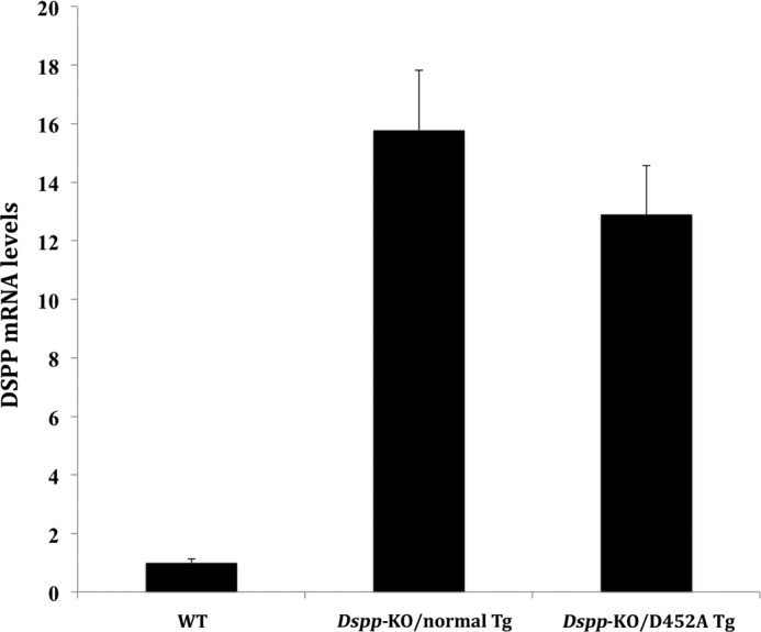
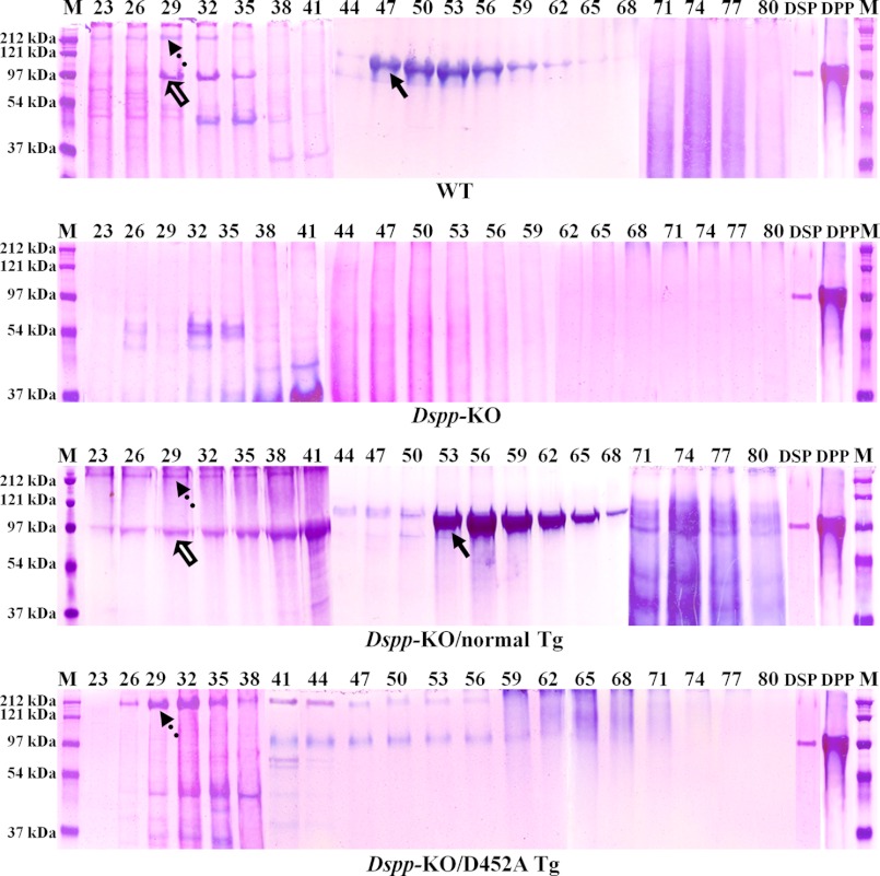
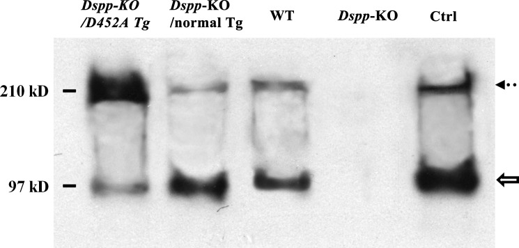
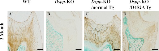
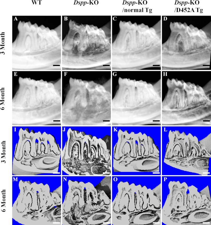
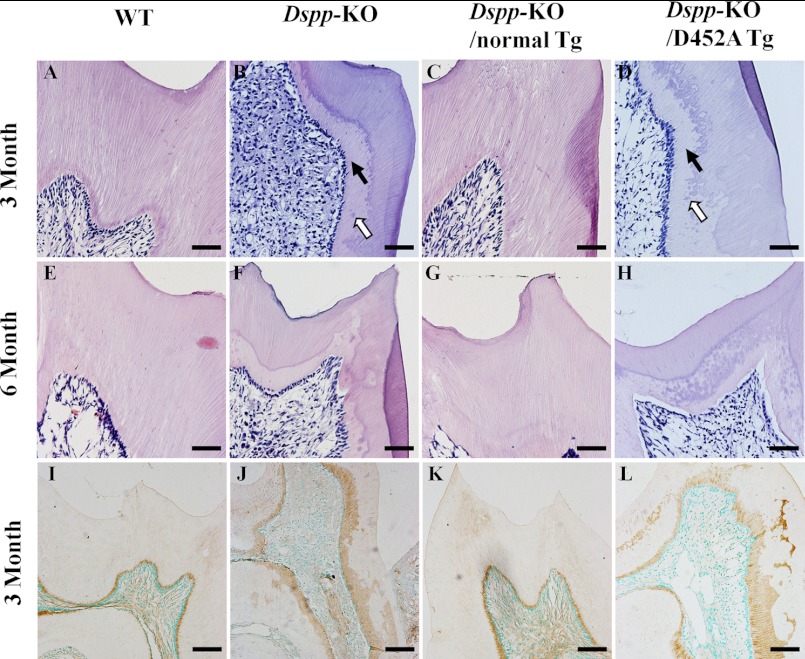
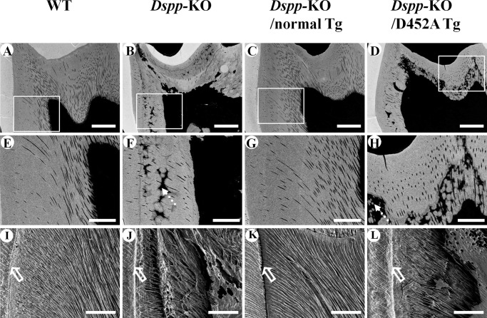
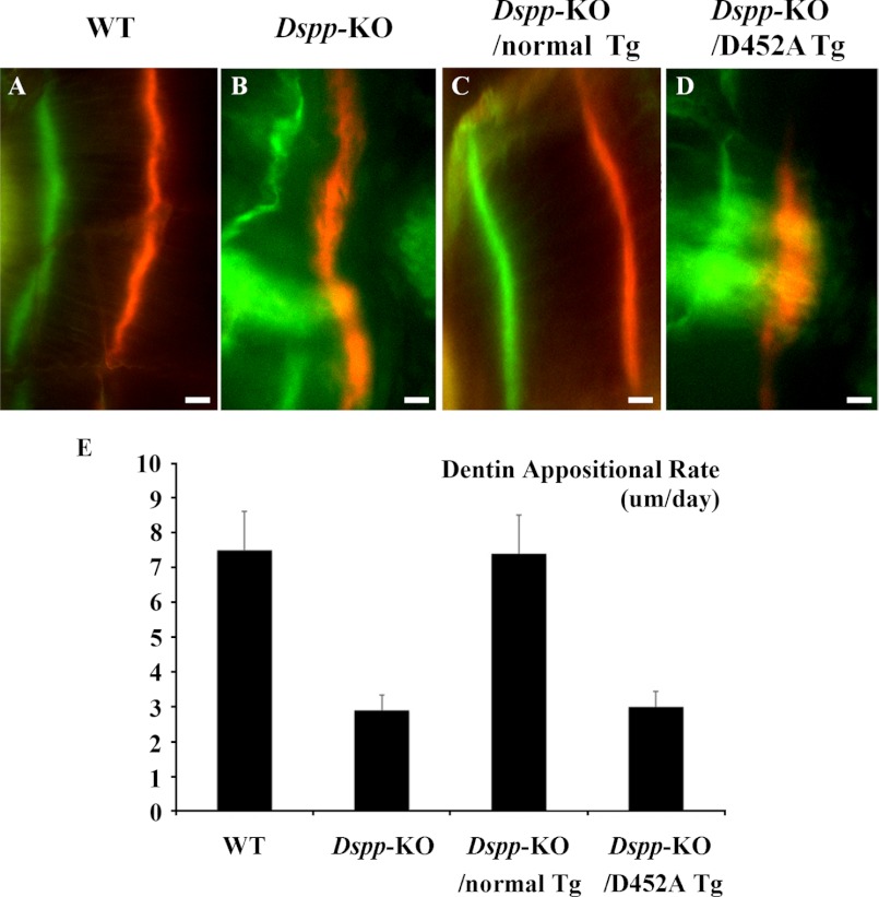
DSPP, which plays a crucial role in dentin formation, is processed into the NH(2)-terminal and COOH-terminal fragments. We believe that the proteolytic processing of DSPP is an essential activation step for its biological function in biomineralization. We tested this hypothesis by analyzing transgenic mice expressing the mutant D452A-DSPP in the Dspp-knock-out (Dspp-KO) background (referred to as "Dspp-KO/D452A-Tg" mice). We employed multipronged approaches to characterize the dentin of the Dspp-KO/D452A-Tg mice, in comparison with Dspp-KO mice and mice expressing the normal DSPP transgene in the Dspp-KO background (named Dspp-KO/normal-Tg mice). Our analyses showed that 90% of the D452A-DSPP in the dentin of Dspp-KO/D452A-Tg mice was not cleaved, indicating that D452A substitution effectively blocked the proteolytic processing of DSPP in vivo. While the expression of the normal DSPP fully rescued the dentin defects of the Dspp-KO mice, expressing the D452A-DSPP failed to do so. These results indicate that the proteolytic processing of DSPP is an activation step essential to its biological function in dentinogenesis.


Figures

FIGURE 1.
FIGURE 1.
DSPP mRNA levels in the incisor of the Dspp-KO/D452A-Tg and Dspp-KO/normal-Tg mice. RNA isolated from the incisor of 1-month-old mice was used for real-time PCR analyses. The mRNA level in the WT mouse incisor was taken as one, while that of the Dspp-KO/normal-Tg or Dspp-KO/D452A-Tg mice was expressed as fold over the WT mice. The level of the transgenic DSPP mRNA in the Dspp-KO/normal-Tg mice was ∼16-fold of that of the endogenous Dspp gene in the WT mice. The level of mRNA from the D452A-DSPP transgene in the Dspp-KO/D452A-Tg mice was ∼13-fold of that of the endogenous Dspp gene in the WT mice. The forward primer sequence used for real-time PCR analysis was from exon 3 of the endogenous Dspp gene, while the reverse was from exon 4. The results were from five analyses (n = 5) for each group.
FIGURE 2.
FIGURE 2.
Stains-All staining of acidic proteins (stained blue or purple) in the NCP extracts from mouse dentin. The NCPs were extracted from the dentin of 3-month-old WT, Dspp-KO, Dspp-KO/normal-Tg and Dspp-KO/D452A-Tg mice. The extracted NCPs were separated into 118 fractions (0.5 ml/fraction) by a Q-Sepharose ion-exchange column; the digits on the top of each image represent the fraction numbers. 60 μl of sample from each of the fractions that potentially contained DSPP-derived products was loaded onto 5–15% SDS-PAGE. The dotted arrows denote DSPP, while the hollow arrows indicate DSP; their identities as DSPP and DSP were confirmed by Western immunoblotting (see Fig. 3). The major blue bands in fractions 47–65 in the WT and Dspp-KO/normal-Tg mice (solid arrows) was primarily made of DPP, although these bands also contained a small amount of bone sialoprotein (BSP), which was confirmed by anti-BSP Western immunoblotting (data not shown). It should be noted that no anti-DPP antibodies are available to detect DPP in Western immunoblotting analyses. Note the abundance of DSP and DPP in the samples from the WT and Dspp-KO/normal-Tg mouse incisors as well as the large amounts of full-length DSPP in the samples from the Dspp-KO/D452A-Tg mice. M, molecular weight standard; DSP, pure DSP isolated from rat dentin; DPP, pure DPP isolated from rat dentin.
FIGURE 3.
FIGURE 3.
Western immunoblotting to detect DSPP and DSP in the NCP extracts from mouse dentin. Western immunoblotting with polyclonal anti-DSP antibodies was used to detect DSPP and DSP in the dentin extracts from the four types of mice. The partially purified rat dentin extract (0.1 μg) containing rat DSPP and DSP was used as a positive control (Ctrl). The Western immunoblotting results from a representative fraction (fraction 29, 60 μl) are shown here. While DSP (hollow arrow) and DSPP (dotted arrow) were detected in the dentin extracts from the Dspp-KO/D452A-Tg, Dspp-KO/normal-Tg, and WT mice, the ratios of DSP to DSPP among these three types of samples were remarkably different. Based on our integrated calculation from triplicate analyses (n = 3) using the image J program, we estimated that the ratio of DSP to DSPP in the Dspp-KO/D452A-Tg mice was 1:10, while that in the Dspp-KO/normal-Tg mice was 15:1. In other words, if the quantity of DSP was used for normalization, the amount of DSPP in the Dspp-KO/D452A-Tg mice would be ∼150 times that of the full-length protein in the Dspp-KO/normal-Tg mice. The findings from both Stains-All and Western immunoblotting analyses showed that the D452A substitution effectively blocked the proteolytic processing of DSPP in the mouse teeth.
FIGURE 4.
FIGURE 4.
Anti-DSP immunohistochemistry. The specimens were from the first mandibular molars of four types of mice at postnatal 3 months. The samples from the Dspp-KO mice (B) were used as negative controls. Anti-DSP activity was observed in the dentin matrix of the WT (A), Dspp-KO/normal-Tg (C), and Dspp-KO/D452A-Tg (D) mice. The signal for the anti-DSP antibody in the dentin of the Dspp-KO/D452A-Tg mice was weaker than in the WT or Dspp-KO/normal-Tg mice. Bar: 50 μm.
FIGURE 5.
FIGURE 5.
Plain x-ray analyses (A–H) and the μ-CT analyses (I–P) of mandibles from 3- and 6-month-old mice. At postnatal 3 months, the WT mice had evenly distributed and well mineralized dentin (A). The mandibular molars in the Dspp-KO mice (B) had an enlarged pulp chamber and thinner dentin compared with the WT mice. The tooth defects in the Dspp-KO/D452A-Tg mice (D) were similar to those of the Dspp-KO mice, whereas the teeth of the Dspp-KO/normal-Tg mice (C) resembled those of the WT mice. At postnatal 6 months, the teeth in the Dspp-KO/normal-Tg mice (G) also appeared the same as those in the WT mice (E), while the teeth of the Dspp-KO/D452A-Tg mice (H) resembled the Dspp-KO mouse teeth (F). In the μ-CT analyses of mandibles, the teeth of the Dspp-KO (J, N) and Dspp-KO/D452A-Tg (L, P) mice had similar dental defects, which included enlarged pulp chambers and thinner dentin. The WT (I, M) and Dspp-KO/normal-Tg (K, O) mice had normal dental structures. Bar: 200 μm.
FIGURE 6.
FIGURE 6.
H&E staining of the dentin-pulp complex in 3- and 6-month-old mice (A–H) and biglycan immunostaining of predentin/dentin in 3-month-old mice (I–L). At postnatal 3 months, the Dspp-KO mice (B) had wider predentin (solid arrow), uncoalescent calcospherites (hollow arrow) and an irregular dentin-predentin border compared with the WT mice (A). The Dspp-KO/D452A-Tg mice (D) showed dentin abnormalities similar to those of the Dspp-KO mice (B). The dentin of the Dspp-KO/normal-Tg (C) mice resembled that of the WT mice (A). At postnatal 6 months, the dentin-pulp structures in the Dspp-KO/normal-Tg (G) mice resembled those of the WT mice (E), while the Dspp-KO/D452A-Tg mouse teeth (H) resembled those of the Dspp-KO mice (F). Bar: A–H = 50 μm. In the biglycan immunostaining of predentin/dentin, the predentin stained by the anti-biglycan antibody (brown color) in the WT mice (I) and Dspp-KO/normal-Tg mice (K) was thin, smooth and evenly distributed, while the predentin in the Dspp-KO mice (J) and Dspp-KO/D452A-Tg mice (L) was wider and unevenly distributed. Bar: I–L = 100 μm.
FIGURE 7.
FIGURE 7.
Backscattered SEM analyses (A–H) and resin infiltration and acid-etched SEM analyses (I–L) of the mandibular first molar from 3-month-old mice. In the backscattered SEM images, the white areas represent the regions with greater amounts of mineral (higher level of mineralization), while the black areas indicate those with less mineral (lower level of mineralization). The dentin in the Dspp-KO (B, F) and Dspp-KO/D452A-Tg mice (D, H) had more black areas (dotted arrows) than in the WT (A, E) or Dspp-KO/normal-Tg (C, G) mice, indicating that the former two had more hypomineralized areas than in the latter two. The images in E–H are enlarged views of the boxed areas in A–D. Bar: A-D = 100 μm, E–H = 40 μm. In the resin infiltration and acid-etched SEM images of the mandibular first molar, the dentinal tubules in the WT mice (I) and Dspp-KO/normal-Tg mice (K) had uniform diameters and were evenly distributed, running parallel to each other and perpendicular to the dental enamel junction (DEJ, hollow arrows). In contrast, the dentinal tubules in the Dspp-KO (J) and Dspp-KO/D452A-Tg (L) mice were tangled, had uneven diameters, and appeared collapsed. Bar: I–L = 50 μm.
FIGURE 8.
FIGURE 8.
Double fluorochrome labeling. The specimens were from the dentin of 5-week-old WT (A), Dspp-KO (B), Dspp-KO/normal-Tg mice (C), and Dspp-KO/D452A-Tg mice (D). In these analyses, the first injection (calcein) produced a green label, while the second injection (Alizarin Red) made a red label. The distance between the green zone and the red zone reflected the width of the dentin matrix that was mineralized in 7 days. Compared with the normal dentin in the WT and Dspp-KO/normal-Tg mice, the labeling zones in the Dspp-KO mice and Dspp-KO/D452A-Tg mice were diffused, indicating an irregular deposition of mineral. Bars = 10 μm. E, quantitative analyses (n = 3) showed that the dentin of the Dspp-KO mice and Dspp-KO/D452A-Tg mice had a remarkably lower mineral deposition rate compared with the WT and Dspp-KO/normal-Tg mice.

Similar articles

Cited by

References

    1. MacDougall M., Simmons D., Luan X., Nydegger J., Feng J., Gu T. T. (1997) Dentin phosphoprotein and dentin sialoprotein are cleavage products expressed from a single transcript coded by a gene on human chromosome 4. Dentin phosphoprotein DNA sequence determination. J. Biol. Chem. 272, 835–842 - PubMed
    1. Butler W. T., Bhown M., Dimuzio M. T., Linde A. (1981) Nonocollagenous proteins of dentin. Isolation and partial characterization of rat dentin proteins and proteoglycans using a three-step preparative method. Coll. Relat. Res. 1, 187–199 - PubMed
    1. Veis A., Perry A. (1967) The phosphoprotein of the dentin matrix. Biochemistry 6, 2409–2416 - PubMed
    1. Xiao S., Yu C., Chou X., Yuan W., Wang Y., Bu L., Fu G., Qian M., Yang J., Shi Y., Hu L., Han B., Wang Z., Huang W., Liu J., Chen Z., Zhao G., Kong X. (2001) Dentinogenesis imperfecta 1 with or without progressive hearing loss is associated with distinct mutations in DSPP. Nat. Genet. 27, 201–204 - PubMed
    1. Zhang X., Zhao J., Li C., Gao S., Qiu C., Liu P., Wu G., Qiang B., Lo W. H., Shen Y. (2001) DSPP mutation in dentinogenesis imperfecta Shields type II. Nat. Genet. 27, 151–152 - PubMed

Publication types