

The acetabular wall index for assessing anteroposterior femoral head coverage in symptomatic patients
- PMID: 22798137
- PMCID: PMC3492620
- DOI: 10.1007/s11999-012-2477-2
The acetabular wall index for assessing anteroposterior femoral head coverage in symptomatic patients
Abstract
Background: Understanding acetabular pathomorphology is necessary to correctly treat patients with hip complaints. Existing radiographic parameters classify acetabular coverage as deficient, normal, or excessive but fail to quantify contributions of anterior and posterior wall coverage. A simple, reproducible, and valid measurement of anterior and posterior wall coverage in patients with hip pain would be a clinically useful tool.
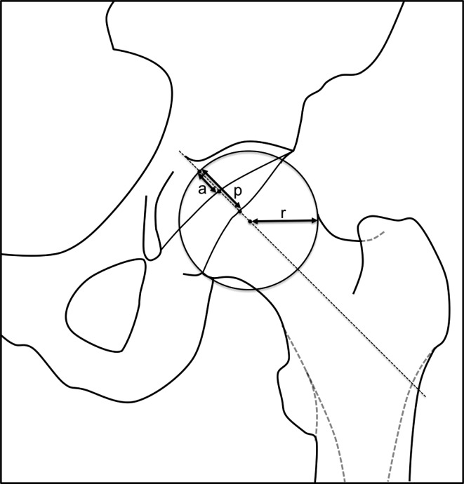
Questions/purposes: We (1) introduce the anterior wall index (AWI) and posterior wall index (PWI), (2) report the intra- and interobserver reliability of these measurements, and (3) validate these measurements against an established computer model.
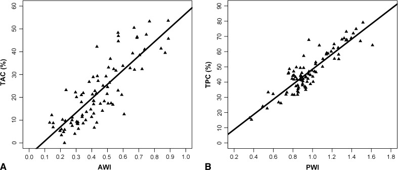
Methods: We retrospectively reviewed 87 hips (63 patients) with symptomatic hip disease. A validated computer model was used to determine total anterior and posterior acetabular coverage (TAC and TPC) on an AP pelvis radiograph. Two independent observers measured the AWI and PWI on each film, and the intraclass correlation coefficient (ICC) was calculated. Pearson correlation was used to determine the strength of linear dependence between our measurements and the computer model.
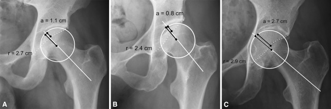
Results: Intra- and interobserver ICCs were 0.94 and 0.99 for the AWI and 0.81 and 0.97 for the PWI. For validation against the computer model, Pearson r values were 0.837 (AWI versus TAC) and 0.895 (PWI versus TPC). Mean AWI and PWI were 0.28 and 0.81 for dysplastic hips, 0.41 and 0.91 for normal hips, 0.61 and 1.15 for hips with a deep acetabulum.
Conclusions: Our data suggest these measures will be helpful in evaluating anterior and posterior coverage before and after surgery but need to be evaluated in asymptomatic individuals without hip abnormalities to establish normal ranges.
Level of evidence: Level III, diagnostic study. See Instructions for Authors for a complete description of levels of evidence.
Figures
References
-
- Chosa E, Tajima N, Nagatsuru Y. Evaluation of acetabular coverage of the femoral head with anteroposterior and false profile radiographs of hip joint. J Orthop Sci. 1997;2:378–390. doi: 10.1007/BF02488925. - DOI
-
- Dutoit M, Zambelli PY. Simplified 3D-evaluation of periacetabular osteotomy. Acta Orthop Belg. 1999;65:288–294. - PubMed
-
- Ecker TM, Tannast M, Puls M, Siebenrock KA, Murphy SB. Pathomorphologic alterations predict presence or absence of hip osteoarthrosis. Clin Orthop Relat Res. 2007;465:46–52. - PubMed
-
- Giori NJ, Trousdale RT. Acetabular retroversion is associated with osteoarthritis of the hip. Clin Orthop Relat Res. 2003;417:263–269. - PubMed
Publication types
MeSH terms
LinkOut - more resources
Full Text Sources
Medical
Research Materials
