Case Reports
. 2010 Dec 1:2010:bcr0920103311.
doi: 10.1136/bcr.09.2010.3311.

Focal nodular hyperplasia with major sinusoidal dilatation: a misleading entity

Affiliations
Case Reports

Focal nodular hyperplasia with major sinusoidal dilatation: a misleading entity

Hervé Laumonier et al. BMJ Case Rep. .

Abstract

Focal nodular hyperplasia (FNH) is a benign liver lesion thought to be a non-specific response to locally increased blood flow. Although the diagnosis of FNH and hepatocellular adenoma (HCA) has made great progress over the last few years using modern imaging techniques, there are still in daily practice some difficulties concerning some atypical nodules. Here, the authors report the case of a 47-year-old woman with a single liver lesion thought to be, by imaging, an inflammatory HCA with major sinusoidal congestion. This nodule was revealed to be, at the microscopical level and after specific immunostaining and molecular analysis, an FNH with sinusoidal dilatation (so-called telangiectatic focal nodular hyperplasia).


Conflict of interest statement

Competing interests None.

Figures

Figure 1
Figure 1
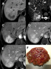
MRI. (A) Axial fat suppressed T1W sequence shows a 55 mm diameter heterogenous nodule in the segment VII. (B) Axial fat suppressed T2W image shows heterogeneity of the lesion with a large hyperintense central area not suggestive of focal nodular hyperplasia (FNH). (C,D,E) Gadolinium enhanced fat suppressed T1W images in the arterial (C), portal venous (D) and delayed (E) phases: strong arterial enhancement of the nodule and heterogenous sustained enhancement in the portal and venous phases. The areas with delayed enhancement are corresponding with the highly hyperintense areas in T2W images (asterisks) suggesting areas of sinusoidal congestion in an inflammatory adenoma. (F) Cut fresh section of the FNH. The gross aspect looks more like a FNH than a hepatocellular adenoma, although there is no stellate scar.
Figure 2
Figure 2
(A) Biopsy specimen performed on the resected nodule at low magnification. (B) H&E staining at high magnification: dilated sinusoids are visible (right) associated with thick walled arteries. This aspect could suggest the diagnosis of hepatocellular adenoma; however, the glutamine synthetase (GS) immunostaining (C) is characteristic of a focal nodular hyperplasia.
Figure 3
Figure 3
In this area, (A) trichrome (TRI) staining and (B) α smooth muscle (SMA) immunostaining, two zones are easily identified: one with dilated sinusoids and one compact. (C,D) Illustrate zones in the vicinity of A and B. Overall, there is little fibrotic bands visible on the trichrome staining.
Figure 4
Figure 4
(A,B,C) are identical zones with different stainings. (A) Heavy glutamine synthetase (GS) staining focalised in the compact zone (right) is faintly present in the dilated zone (left); in this zone, dilated sinusoids surround areas containing many arteries (B) and cytokeratin 7 (CK7) positive cells (C,D) mixed with inflammatory cells. SMA, α smooth muscle.
Figure 5
Figure 5
Compact zone. (A) Illustrates the typical aspect of glutamine synthetase (GS) in focal nodular hyperplasia (compact zone). This staining is sharply different in the non-tumoural liver (NTL) where the staining is limited around the hepatic veins. (C) Numerous arteries are visible in the vicinity of veins surrounded by areas that highly express GS (D). SMA, α smooth muscle.
Figure 6
Figure 6
Compact zone. (A,B,C,D) Illustrate the contact between arteries and veins. SMA, α smooth muscle.
Figure 7
Figure 7
Compact zone. The veins in (A) and (B) are occlude and in part recanalised. (C,D) The lumen of this artery surrounded by connective tissue is not visible. SMA, α smooth muscle. TRI, trichrome.

Similar articles

Cited by

References

    1. Wanless IR, Albrecht S, Bilbao J, et al. Multiple focal nodular hyperplasia of the liver associated with vascular malformations of various organs and neoplasia of the brain: a new syndrome. Mod Pathol 1989;2:456–62 - PubMed
    1. Nguyen BN, Fléjou JF, Terris B, et al. Focal nodular hyperplasia of the liver: a comprehensive pathologic study of 305 lesions and recognition of new histologic forms. Am J Surg Pathol 1999;23:1441–54 - PubMed
    1. Paradis V, Benzekri A, Dargère D, et al. Telangiectatic focal nodular hyperplasia: a variant of hepatocellular adenoma. Gastroenterology 2004;126:1323–9 - PubMed
    1. Bioulac-Sage P, Rebouissou S, Sa Cunha A, et al. Clinical, morphologic, and molecular features defining so-called telangiectatic focal nodular hyperplasias of the liver. Gastroenterology 2005;128:1211–18 - PubMed
    1. Bioulac-Sage P, Rebouissou S, Thomas C, et al. Hepatocellular adenoma subtype classification using molecular markers and immunohistochemistry. Hepatology 2007;46:740–8 - PubMed

Publication types

LinkOut - more resources