. 2012 Sep;153(9):4328-35.
doi: 10.1210/en.2012-1241. Epub 2012 Jul 13.

The potassium channel, Kir3.4 participates in angiotensin II-stimulated aldosterone production by a human adrenocortical cell line

Affiliations

The potassium channel, Kir3.4 participates in angiotensin II-stimulated aldosterone production by a human adrenocortical cell line

Kenji Oki et al. Endocrinology. 2012 Sep.

Abstract

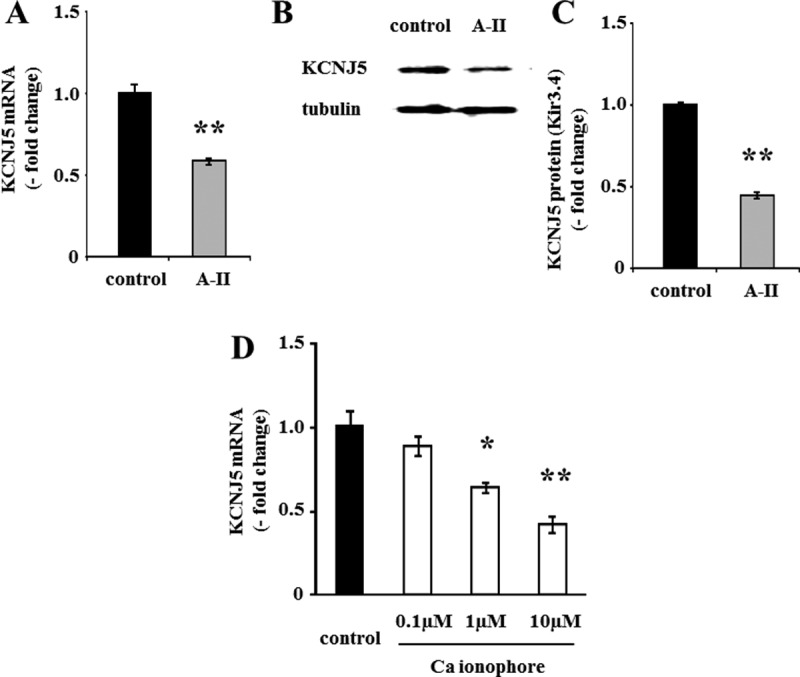
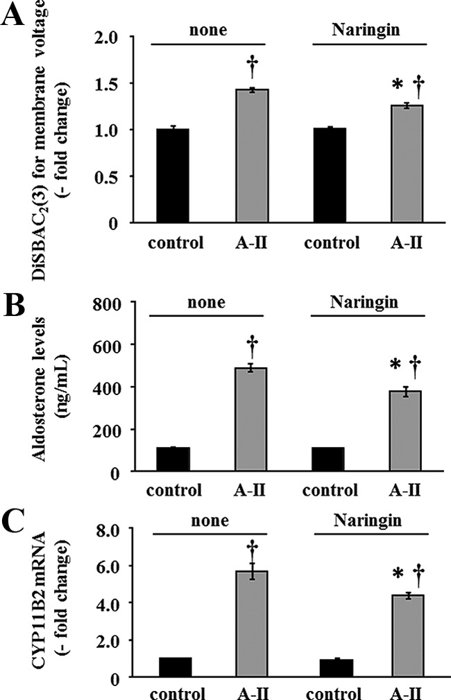
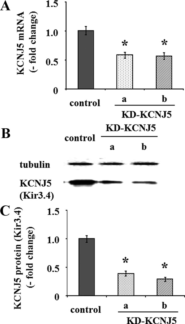
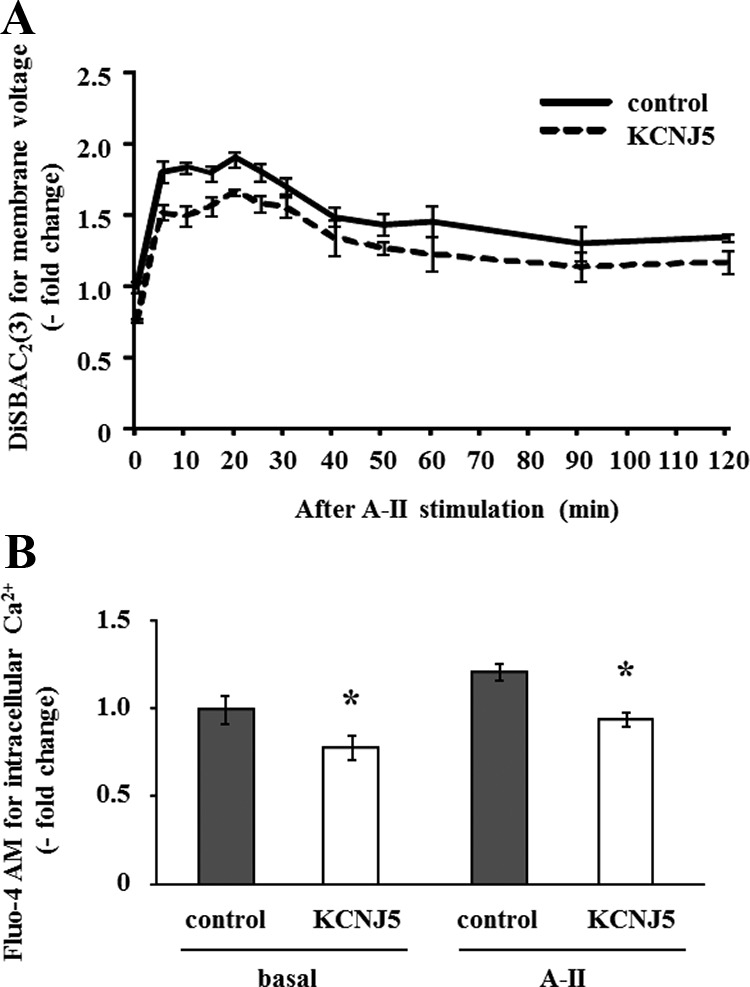
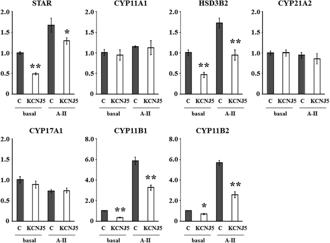
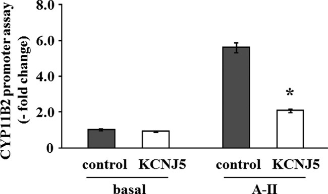
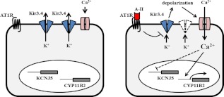
Angiotensin II (A-II) regulation of aldosterone secretion is initiated by inducing cell membrane depolarization, thereby increasing intracellular calcium and activating the calcium calmodulin/calmodulin kinase cascade. Mutations in the selectivity filter of the KCNJ5 gene coding for inward rectifying potassium channel (Kir)3.4 has been found in about one third of aldosterone-producing adenomas. These mutations result in loss of selectivity of the inward rectifying current for potassium, which causes membrane depolarization and opening of calcium channels and activation of the calcium calmodulin/calmodulin kinase cascade and results in an increase in aldosterone secretion. In this study we show that A-II and a calcium ionophore down-regulate the expression of KCNJ5 mRNA and protein. Activation of Kir3.4 by naringin inhibits A-II-stimulated membrane voltage and aldosterone secretion. Overexpression of KCNJ5 in the HAC15 cells using a lentivirus resulted in a decrease in membrane voltage, intracellular calcium, expression of steroidogenic acute regulatory protein, 3-β-hydroxysteroid dehydrogenase 3B2, cytochrome P450 11B1 and cytochrome P450 11B2 mRNA, and aldosterone synthesis. In conclusion, A-II appears to stimulate aldosterone secretion by depolarizing the membrane acting in part through the regulation of the expression and activity of Kir3.4.


Figures

Fig. 1.
Fig. 1.
Effect of A-II and calcium ionophore on mRNA and protein expression of KCNJ5. A and D, Confluent cells were serum deprived in DMEM-F12 containing 0.1% cosmic calf serum for 24 h and then incubated with fresh media with 0.1% serum and no secretagogue, A-II (10 nm), or calcium ionophore A23187 for 3 h. After aspiration of media, the cells were collected for RNA extraction and real-time RT-PCR was performed. Results were normalized by GAPDH mRNA expression and expressed as fold change vs. control. B and C, Cells were serum deprived 72 h after infection in DMEM-F12 containing 0.1% cosmic calf serum for 24 h and then incubated with fresh media with 0.1% serum and no secretagogue or A-II (10 nm) for 8 h. After aspiration of media, the cells were harvested with protease inhibitor. The cell lysates were subjected to immunoblotting analysis using anti-KCNJ5 or antitubulin antibodies. The KCNJ5 band intensity of scanned images was adjusted by that of tubulin and expressed as fold change vs. control. *, P < 0.05 vs. control, n = 3; **, P < 0.01 vs. control, n = 3.
Fig. 2.
Fig. 2.
Effect of naringin on membrane voltage, CYP11B2 mRNA expression, and aldosterone production. Confluent cells were serum deprived in DMEM-F12 containing 0.1% cosmic calf serum for 24 h. Pretreatments with or without naringin for 30 min were performed for each experiment before adding secretagogue. A, HAC15 cells were incubated in HEPES buffer with 3 μm DiSBAC2(3) and no secretagogue or A-II (10 nm) for 5 min. Fluorescence was detected by a plate reader. Results were expressed as fold change vs. control cells. *, P < 0.05 comparison between with and without naringin after A-II stimulation, n = 4. †, P < 0.01 comparison with the respective control, n = 4. B, Cells were incubated with no secretagogue or with A-II (10 nm) and without and with naringin for 6 h, and aldosterone was measured in the media. *, P < 0.05 comparison between with and without naringin after A-II stimulation, n = 4. †, P < 0.01 comparison with the respective control, n = 4. C, After 3 h incubation with no secretagogue or A-II (10 nm) and without and with naringin, cells were harvested for RNA extraction, and real-time RT-PCR was performed. Results were normalized by GAPDH mRNA expression and expressed as fold change vs. control. *, P < 0.05 comparison between with and without naringin after A-II stimulation, n = 4. †, P < 0.01 comparison with the respective control, n = 4.
Fig. 3.
Fig. 3.
Effect of shRNA-KCNJ5 on mRNA and protein expression of KCNJ5. Cells were serum deprived 72 h after transduction with control or either shRNA-KCNJ5 lentiviruses as in Fig 1. A, After aspiration of media, the cells were collected for RNA as in Fig 1. B and C, After aspiration of media, the cells were harvested with protease inhibitor. The cell lysates were subjected to immunoblotting analysis using anti-KCNJ5 or antitubulin antibodies. The KCNJ5 band intensity of scanned images was adjusted by that of tubulin and expressed as fold change vs. control. *, P < 0.01 vs. control, n = 3.
Fig. 4.
Fig. 4.
Effect of the KCNJ5 overexpression on membrane voltage and intracellular Ca2+ concentration in HAC15 cells transduced with control or KCNJ5 lentiviruses. Cells were serum deprived 72 h after infection in DMEM-F12 containing 0.1% cosmic calf serum for 24 h, and then incubated in HEPES buffer with 10 nm A-II and 3 μm DiSBAC2(3) for the indicated time (A) or 3 μm Fluo-4 AM for 10 min (B). Fluorescence was detected by a plate reader. Results were expressed as fold change vs. control cells. *, P < 0.05 vs. control, n = 4.
Fig. 5.
Fig. 5.
Basal and A-II-stimulated aldosterone and cortisol production by HAC15 transduced with control, shRNA-KCNJ5 (A), or KCNJ5 lentiviruses (B, C). Cells were serum deprived 72 h after infection in DMEM-F12 containing 0.1% cosmic calf serum for 24 h after which cells were incubated with no secretagogue or A-II (10 nm) for 24 h. *, P < 0.05 vs. control, n = 4, **, P < 0.01 vs. control, n = 4.
Fig. 6.
Fig. 6.
Effect of the KCNJ5 overexpression on mRNA expression of the key proteins for adrenal steroid synthesis such as StAR, CYP11A1, HSD3B2, CYP21A2, CYP17A1, CYP11B1, and CYP11B2. Cells were serum deprived 72 h after infection in DMEM-F12 containing 0.1% cosmic calf serum for 24 h and then incubated with fresh media with 0.1% serum for 3 h. After aspiration of media, the cells were collected for RNA extraction and real-time RT-PCR was performed. Results were normalized by GAPDH mRNA expression and expressed as fold change vs. control. *, P < 0.05 vs. control, n = 3, **, P < 0.01 vs. control, n = 3.
Fig. 7.
Fig. 7.
Effect of the KCNJ5 overexpression on CYP11B2 reporter gene expression. HAC15 cells stably infected with pBM14-CYP11B2 promoter/gaussia were infected with control or KCNJ5 lentiviruses. After 72 h incubation, cells were serum deprived in DMEM-F12 containing 0.1% cosmic calf serum for 24 h and incubated in fresh media with 0.1% serum for 24 h, and supernatants were collected to assess luciferase activity. Results are shown as fold change vs. control cells. *, P < 0.01 vs. control, n = 3.
Fig. 8.
Fig. 8.
A possible mechanism by which A-II regulates aldosterone production via Kir3.4. A-II stimulates aldosterone synthesis (CYP11B2) by increasing intracellular Ca2+ concentration through inhibition of Kir3.4 receptor and activation of voltage-gated Ca2+ channel. The increase of intracellular Ca2+ suppresses Kir3.4 expression.

References

    1. Funder JW, Carey RM, Fardella C, Gomez-Sanchez CE, Mantero F, Stowasser M, Young WF, Jr, Montori VM. 2008. Case detection, diagnosis, and treatment of patients with primary aldosteronism: an Endocrine Society clinical practice guideline. J Clin Endocrinol Metab 93:3266–3281 - PubMed
    1. Milliez P, Girerd X, Plouin PF, Blacher J, Safar ME, Mourad JJ. 2005. Evidence for an increased rate of cardiovascular events in patients with primary aldosteronism. J Am Coll Cardiol 45:1243–1248 - PubMed
    1. Condon JC, Pezzi V, Drummond BM, Yin S, Rainey WE. 2002. Calmodulin-dependent kinase I regulates adrenal cell expression of aldosterone synthase. Endocrinology 143:3651–3657 - PubMed
    1. Otis M, Gallo-Payet N. 2006. Differential involvement of cytoskeleton and rho-guanosine 5′-triphosphatases in growth-promoting effects of angiotensin II in rat adrenal glomerulosa cells. Endocrinology 147:5460–5469 - PubMed
    1. Nogueira EF, Rainey WE. 2010. Regulation of aldosterone synthase by activator transcription factor/cAMP response element-binding protein family members. Endocrinology 151:1060–1070 - PMC - PubMed

Publication types

MeSH terms