

Group I p21-activated kinases (PAKs) promote tumor cell proliferation and survival through the AKT1 and Raf-MAPK pathways
- PMID: 22798428
- PMCID: PMC3447107
- DOI: 10.1158/1541-7786.MCR-12-0082
Group I p21-activated kinases (PAKs) promote tumor cell proliferation and survival through the AKT1 and Raf-MAPK pathways
Abstract
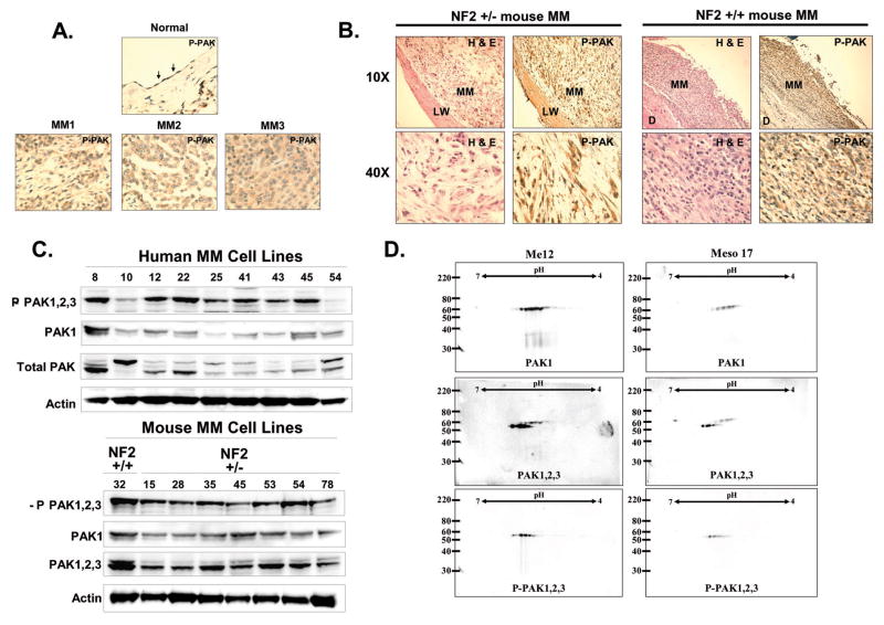
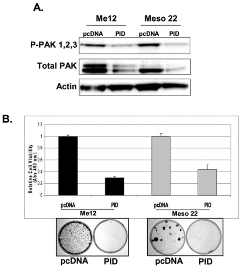
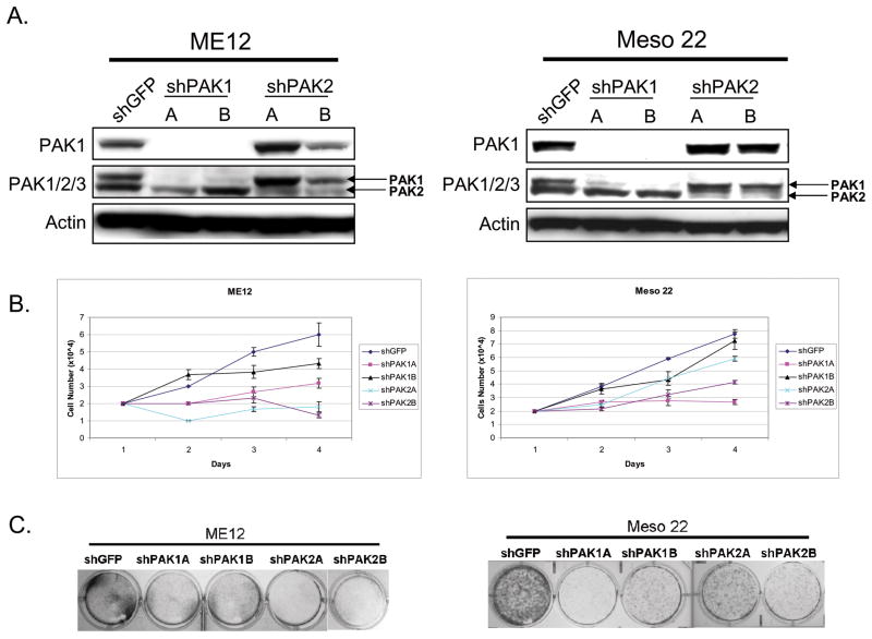
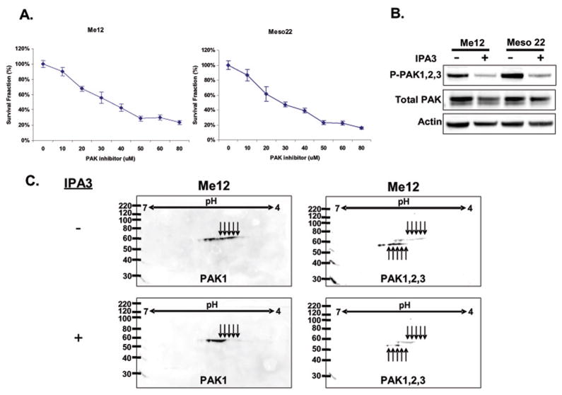
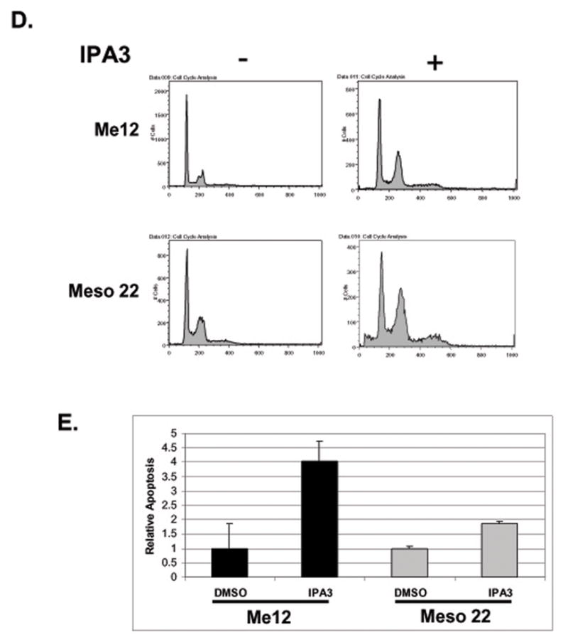
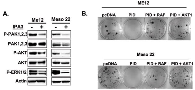
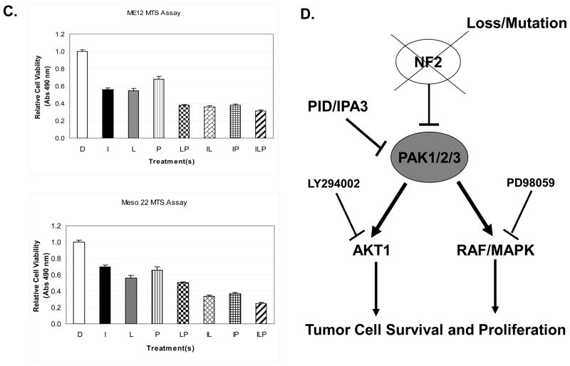
Group I p21-activated kinases (PAK) are important effectors of the small GTPases Rac and Cdc42, which regulate cell motility/migration, survival, proliferation, and gene transcription. Hyperactivation of these kinases have been reported in many tumor types, making PAKs attractive targets for therapeutic intervention. PAKs are activated by growth factor-mediated signaling and are negatively regulated by the tumor suppressor neurofibromatosis type 2 (NF2)/Merlin. Thus, tumors characterized by NF2 inactivation would be expected to show hyperactivated PAK signaling. On the basis of this rationale, we evaluated the status of PAK signaling in malignant mesothelioma, an aggressive neoplasm that is resistant to current therapies and shows frequent inactivation of NF2. We show that group I PAKs are activated in most mesotheliomas and mesothelioma cell lines and that genetic or pharmacologic inhibition of PAKs is sufficient to inhibit mesothelioma cell proliferation and survival. We also identify downstream effectors and signaling pathways that may contribute mechanistically to PAK-related tumorigenesis. Specifically, we show that inhibition of PAK results in attenuation of AKT and Raf-MAPK signaling and decreased tumor cell viability. Collectively, these data suggest that pharmacologic inhibition of group I PAKs may have therapeutic efficacy in tumors characterized by PAK activation.
Conflict of interest statement
No potential conflicts of interest were disclosed.
Figures
Similar articles
-
p21-Activated kinases are required for transformation in a cell-based model of neurofibromatosis type 2.PLoS One. 2010 Nov 2;5(11):e13791. doi: 10.1371/journal.pone.0013791. PLoS One. 2010. PMID: 21072183 Free PMC article.
-
Group I Paks as therapeutic targets in NF2-deficient meningioma.Oncotarget. 2015 Feb 10;6(4):1981-94. doi: 10.18632/oncotarget.2810. Oncotarget. 2015. PMID: 25596744 Free PMC article.
-
FRAX597, a small molecule inhibitor of the p21-activated kinases, inhibits tumorigenesis of neurofibromatosis type 2 (NF2)-associated Schwannomas.J Biol Chem. 2013 Oct 4;288(40):29105-14. doi: 10.1074/jbc.M113.510933. Epub 2013 Aug 19. J Biol Chem. 2013. PMID: 23960073 Free PMC article.
-
P21 activated kinase signaling in cancer.Semin Cancer Biol. 2019 Feb;54:40-49. doi: 10.1016/j.semcancer.2018.01.006. Epub 2018 Jan 9. Semin Cancer Biol. 2019. PMID: 29330094 Review.
-
NF2/Merlin Inactivation and Potential Therapeutic Targets in Mesothelioma.Int J Mol Sci. 2018 Mar 26;19(4):988. doi: 10.3390/ijms19040988. Int J Mol Sci. 2018. PMID: 29587439 Free PMC article. Review.
Cited by
-
Multifaceted p21 in carcinogenesis, stemness of tumor and tumor therapy.World J Stem Cells. 2020 Jun 26;12(6):481-487. doi: 10.4252/wjsc.v12.i6.481. World J Stem Cells. 2020. PMID: 32742565 Free PMC article. Review.
-
microRNA-126 suppresses PAK4 expression in ovarian cancer SKOV3 cells.Oncol Lett. 2015 May;9(5):2225-2229. doi: 10.3892/ol.2015.3012. Epub 2015 Mar 3. Oncol Lett. 2015. PMID: 26137045 Free PMC article.
-
IGF1R signaling drives antiestrogen resistance through PAK2/PIX activation in luminal breast cancer.Oncogene. 2018 Apr;37(14):1869-1884. doi: 10.1038/s41388-017-0027-9. Epub 2018 Jan 22. Oncogene. 2018. PMID: 29353882
-
Deciphering the link between PI3K and PAK: An opportunity to target key pathways in pancreatic cancer?Oncotarget. 2017 Feb 21;8(8):14173-14191. doi: 10.18632/oncotarget.13309. Oncotarget. 2017. PMID: 27845911 Free PMC article. Review.
-
Pak2 restrains endomitosis during megakaryopoiesis and alters cytoskeleton organization.Blood. 2015 May 7;125(19):2995-3005. doi: 10.1182/blood-2014-10-604504. Epub 2015 Mar 30. Blood. 2015. PMID: 25824689 Free PMC article.
References
-
- Xiao GH, Beeser A, Chernoff J, Testa JR. p21-activated kinase links Rac/Cdc42 signaling to merlin. J Biol Chem. 2002;277:883–6. - PubMed
-
- Eswaran J, Soundararajan M, Knapp S. Targeting group II PAKs in cancer and metastasis. Cancer Metastasis Rev. 2009;28:209–17. - PubMed
-
- Eswaran J, Soundararajan M, Kumar R, Knapp S. UnPAKing the class differences among p21-activated kinases. Trends Biochem Sci. 2008;33:394–403. - PubMed
Publication types
MeSH terms
Substances
Grants and funding
LinkOut - more resources
Full Text Sources
Medical
Molecular Biology Databases
Research Materials
Miscellaneous
