. 2012 Nov;10(11):1440-50.
doi: 10.1158/1541-7786.MCR-12-0158. Epub 2012 Jul 13.

Functional effects of GRM1 suppression in human melanoma cells

Affiliations

Functional effects of GRM1 suppression in human melanoma cells

Janet Wangari-Talbot et al. Mol Cancer Res. 2012 Nov.

Abstract

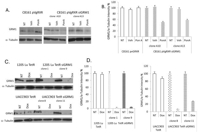
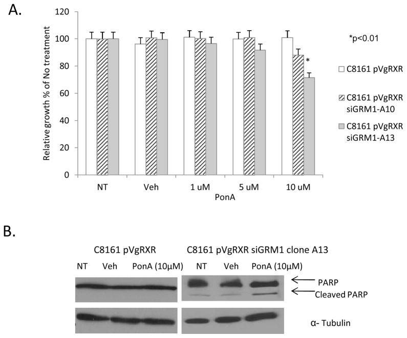
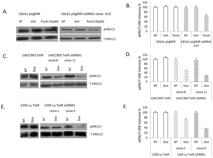
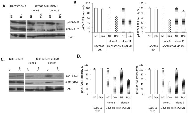
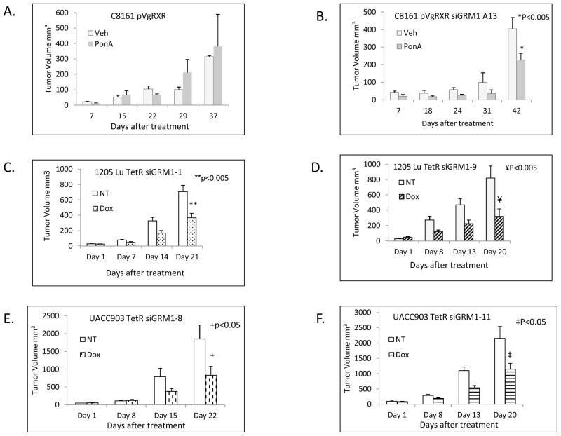
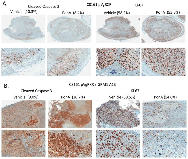
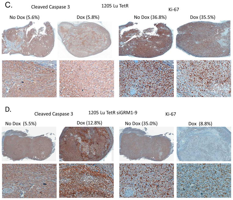
Ectopic expression of a neuronal receptor, metabotropic glutamate receptor 1 (Grm1), in melanocytes has been implicated in melanoma development in mouse models. The human relevance of this receptor's involvement in melanoma pathogenesis was shown by detecting GRM1 expression in subsets of human melanomas, an observation lacking in benign nevi or normal melanocytes. Grm1-transformed mouse melanocytes and a conditional Grm1 transgenic mouse model confirmed a requirement for sustained expression of Grm1 for the maintenance of transformed phenotypes in vitro and tumorigenicity in vivo. Here, we investigate if continued GRM1 expression is also required in human melanoma cell lines by using two inducible, silencing RNA systems: the ecdysone/Ponasterone A and tetracycline on/off approaches to regulate GRM1 expression in the presence of each inducer. Various in vitro assays were conducted to assess the consequences of a reduction in GRM1 expression on cell proliferation, apoptosis, downstream targeted signaling pathways, and in vivo tumorigenesis. We showed that suppression of GRM1 expression in several human melanoma cell lines resulted in a reduction in the number of viable cells and a decrease in stimulated mitogen-activated protein kinase (MAPK) and PI3K/AKT and suppressed tumor progression in vivo. These results reinforce earlier observations where a reduction in cell growth in vitro and tumorigenesis in vivo were correlated with decreased GRM1 activities by pharmacologic inhibitors of the receptor, supporting the notion that GRM1 plays a role in the maintenance of transformed phenotypes in human melanoma cells in vitro and in vivo and could be a potential therapeutic target for the treatment of melanoma.


Conflict of interest statement

Conflict of interest: None disclosed

Figures

Figure 1
Figure 1. Inducible suppression of GRM1
(A.) Suppression of GRM1 protein expression in the ecdysone regulated system in C8161 human melanoma cells in the presence of the inducer, 10 μM Ponasterone A (PonA) in two independent clones. No suppression is observed in control C8161 pVgRXR cells in the presence of PonA. Samples were normalized to α-Tubulin. NT- No treatment, Veh - (Vehicle) DMSO. (B.) Quantitation of C8161 pVgRXR siGRM1 clones A10 and A13 western blots from A. using Optiquant software to determine % change in intensity. (C.) Suppression of GRM1 expression using the tetracycline/doxycycline regulated system in two independent clones from 1205 Lu and UACC903 human melanoma cell lines in the presence of 10 ng/ml doxycycline. No suppression is observed in control 1205 Lu TetR and UACC903 TetR in the presence of doxycycline. Equal loading was shown by levels of α-Tubulin. NT-No treatment, Dox- Doxycycline. (D.) Quantitation of western blots of 1205 Lu TetR siGRM1 and UACC903 TetR siGRM1 clones from panel C using Optiquant software to determine % change in intensity.
Figure 2
Figure 2. In vitro growth properties in C8161 siGRM1 human melanoma cells
(A.) MTT cell viability/proliferation assays showing decreased viability and proliferation in C8161 human melanoma cells with suppressed GRM1 expression by the ecdysone regulated system in the presence of 10 μM PonA. (B.) C8161 pVgRXR siGRM1 cells treated with 10 μM PonA showing increased cleavage of PARP an indicator of apoptosis but not in control C8161 pVgRXR cells. Equal loading was shown by levels of α-Tubulin. NT - No treatment, Veh - Vehicle- DMSO, PonA - Ponasterone A.
Figure 3
Figure 3. Modulation of MAPK signaling with GRM1 siRNA
(A.) Western immunoblots of protein lysates prepared from C8161 pVgRXR or C8161 pVgRXR siGRM1-A13 cells, not treated (NT) or treated with vehicle (DMSO) or with PonA and probed for phosphorylated (pERK1/2) and total ERK1/2 (T-ERK1/2). (B) Quantification of the intensities on the immunoblots from A. with Optiquant software with NT or Veh set as 100%. (C.) Suppression of ERK1/2 phosphorylation in UACC903 TetR siGRM1 clones but not in control UACC903 TetR after doxycycline (10 ng/ml) treatment. Equal loading was assessed with levels of total ERK1/2. (D.) Quantification of the intensities on the immunoblots from C. with Optiquant software. (E.) Suppression of ERK1/2 phosphorylation in 1205 Lu TetR siGRM1 clones and not in 1205 Lu TetR controls after doxycycline (10 ng/ml treatment). Equal loading was shown by levels of total ERK1/2. NT- No treatment, Dox - doxycycline. (F.) Quantification of the intensities on the immunoblots from E. with Optiquant software.
Figure 4
Figure 4. Modulation of PI3K/AKT signaling by GRM1 siRNA
(A.) Suppression of total AKT (Serine 473) and AKT2 phosphorylation in UACC903 TetR siGRM1 clones 8 and 11, but not in UACC903 TetR after doxycycline (10 ng/ml) treatment. (B.) Quantification of the intensities on the immunoblots from A. with Optiquant software. (C.) Suppression of AKT (Serine 473) and AKT2 phosphorylation in 1205 Lu TetR siGRM1 clones and not in 1205 Lu TetR controls after doxycycline (10 ng/ml) treatment. (D.) Quantification of the intensities on the immunoblots from C. with Optiquant software. Total AKT was used to show equal loading. NT- No treatment, Dox -doxycycline.
Figure 5
Figure 5. In vivo xenograft tumorigenicity assays
(A.) In vivo tumorigenicity assay of C8161 pVgRXR cells or (B.) C8161 pVgRXR siGRM1 A13 cells after treatment with vehicle (olive oil) or PonA (10 mg/kg) administered via intraperitoneal injection. All tumor bearing mice were euthanized after 37–42 days due to tumor burden in the control groups. *p<0.005 when PonA treated C8161 pVgRXR siGRM1 A13 tumor volumes were compared to vehicle treated ones. (C and D.) In vivo tumorigenicity assays of 1205 Lu TetR siGRM1 clone 1 (C) and clone 9 (D) No treatment (NT) or doxycycline treated (0.1% w/v) (Dox). * p<0.005 when 1205 Lu TetR siGRM1-9 Dox treated tumor volumes were compared to the not treated controls at day 21. ** p<0.005 when 1205 Lu TetR siGRM1-1 Dox treated tumor volumes were compared to the not treated controls at day 20. (E and F.) In vivo tumorigenicity assays of UACC903 TetR siGRM1 clone 8 (E) and clone 11 (F). No treatment (NT) and doxycycline treated (0.1% w/v) (Dox). * p<0.05 when UACC903 TetR siGRM1-8 Dox treated tumor volumes were compared to the not treated controls at day 22. ** p<0.05 when UACC903 TetR siGRM1-11 Dox treated tumor volumes were compared to the not treated controls at day 20.
Figure 6
Figure 6. Immunohistochemical (IHC) analysis
IHC performed on excised tumor xenografts from (A) C8161 pVgRXR, (B) C8161 pVgRXR siGRM1 A13 PonA and vehicle treated controls (C) 1205 Lu TetR and (D) 1205 Lu TetR siGRM1-9 no treatment or Dox treated for cleaved Caspase-3 and Ki-67. Top panels, 10X magnification, bottom panels 100X magnifications of a section of the stained tumors. The numbers of stained cells were quantified with a digital Aperio ScanScopeGL system and ImageScope software and are expressed as percentages of the total number of cells counted. When vehicle treated samples are compared with PonA treated samples; cleaved Caspase-3, p<0.05 and Ki-67, p<0.001. When no Dox samples are compared with Dox treated samples; cleaved Caspase-3, p<0.05 and Ki-67, p<0.001.

References

    1. American Cancer Society. Cancer facts & figures. Atlanta: American Cancer Society; 2012.
    1. Zhu H, Reuhl K, Botha R, Ryan K, Wei J, Chen S. Development of early melanocytic lesions in transgenic mice predisposed to melanoma. Pigment Cell Res. 2000;13:158–64. - PubMed
    1. Pollock PM, Cohen-Solal K, Sood R, et al. Melanoma mouse model implicates metabotropic glutamate signaling in melanocytic neoplasia. Nat Genet. 2003;34:108–12. - PubMed
    1. Conn PJ, Pin J-P. Pharmacology and functions of metabotropic glutamate receptors. Annu Rev Pharmacol Toxicol. 1997;37:205–37. - PubMed
    1. Funusaka Y, Harada T, Aiba A, Nishigori C. Expression of metabotropic glutamate receptor 1 and phosphorylated extracellular signal-regulated kinase 1/2 proteins in human melanocytic lesions. Pigm Cell Res. 2006;19:256.

Publication types

MeSH terms