. 2012 Nov;47(5):637-44.
doi: 10.1165/rcmb.2012-0009OC. Epub 2012 Jul 12.

Glucocorticoid receptor and histone deacetylase-2 mediate dexamethasone-induced repression of MUC5AC gene expression

Affiliations

Glucocorticoid receptor and histone deacetylase-2 mediate dexamethasone-induced repression of MUC5AC gene expression

Yajun Chen et al. Am J Respir Cell Mol Biol. 2012 Nov.

Abstract

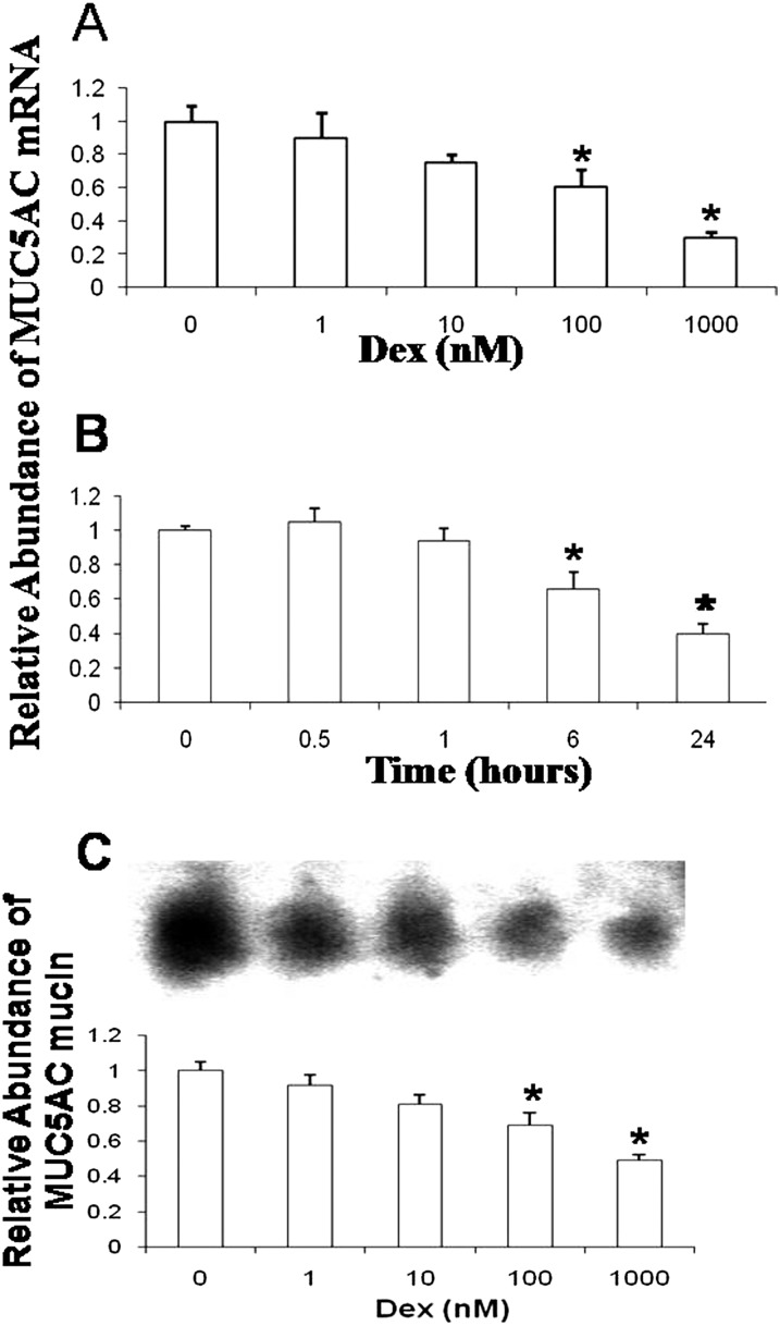
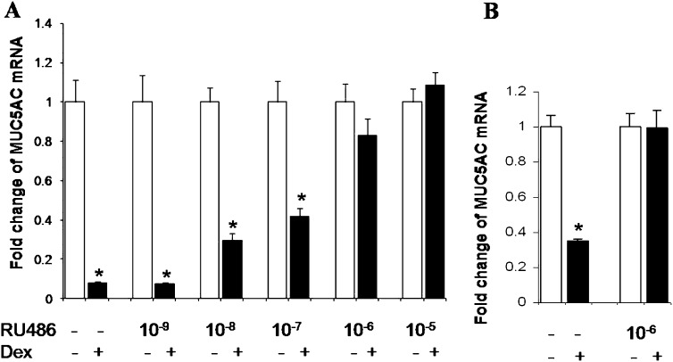
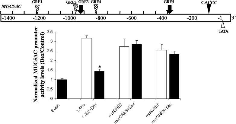
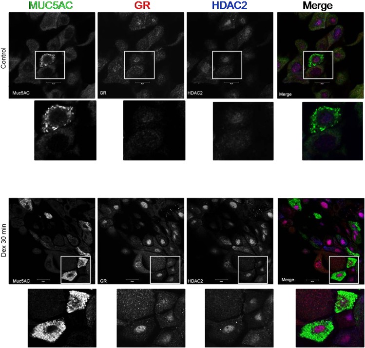
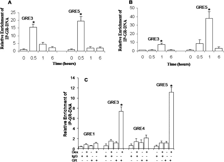
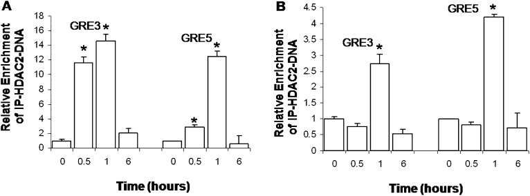
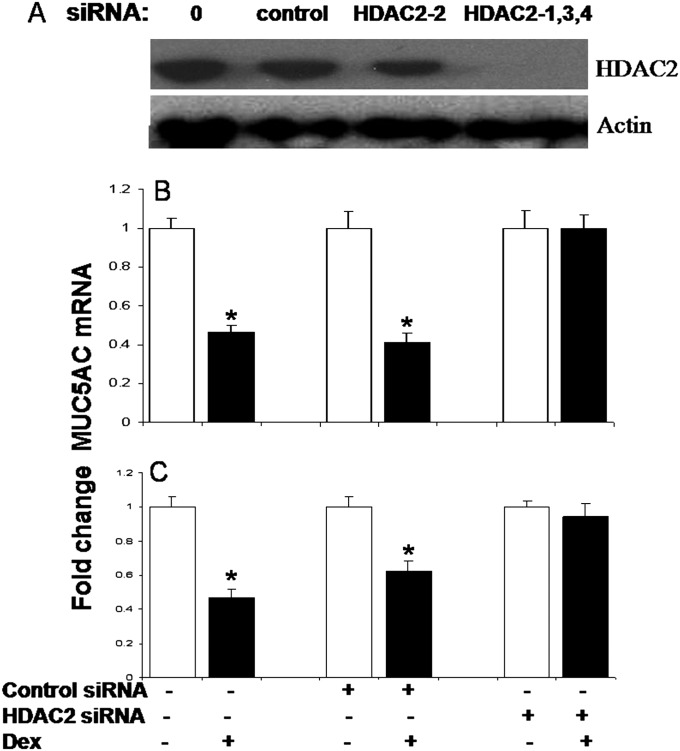
Airway occlusion in obstructive airway diseases is caused in part by the overproduction of secretory mucin glycoproteins through the up-regulation of mucin (MUC) genes by inflammatory mediators. Some pharmacological agents, including the glucocorticoid dexamethasone (Dex), repress mucin concentrations in lung epithelial cancer cells. Here, we show that Dex reduces the expression of MUC5AC, a major airway mucin gene, in primary differentiated normal human bronchial epithelial (NHBE) cells in a dose-dependent and time-dependent manner, and that the Dex-induced repression is mediated by the glucocorticoid receptor (GR) and two glucocorticoid response elements (GREs) in the MUC5AC promoter. The pre-exposure of cells to RU486, a GR antagonist, and mutations in either the GRE3 or GRE5 cis-sites abolished the Dex-induced repression. Chromatin immunoprecipitation (ChIP) assays showed a rapid temporal recruitment of GR to the GRE3 and GRE5 cis-elements in the MUC5AC promoter in NHBE and in A549 cells. Immunofluorescence showed nuclear colocalization of GR and histone deacetylase-2 (HDAC2) in MUC5AC-expressing NHBE cells. ChIP also showed a rapid temporal recruitment of HDAC2 to the GRE3 and GRE5 cis-elements in the MUC5AC promoter in both cell types. The knockdown of HDAC2 by HDAC2-specific short interfering RNA prevented the Dex-induced repression of MUC5AC in NHBE and A549 cells. These data demonstrate that GR and HDAC2 are recruited to the GRE3 and GRE5 cis-sites in the MUC5AC promoter and mediate the Dex-induced cis repression of MUC5AC gene expression. A better understanding of the mechanisms whereby glucocorticoids repress MUC5AC gene expression may be useful in formulating therapeutic interventions in chronic lung diseases.


Figures

Figure 1.
Figure 1.
Dexamethasone (Dex) decreases mucin 5AC (MUC5AC) mRNA and protein concentrations in primary differentiated normal human bronchial epithelial (NHBE) cells in a dose-dependent and time-dependent manner. (A) Cells were exposed to Dex (1, 10, 100, or 1,000 nM) or vehicle for 24 hours. (B) Cells were incubated with 1,000 nM Dex or vehicle for 0, 0.5, 1, 6, and 24 hours. In A and B, MUC5AC and actin mRNA concentrations were quantified by quantitative RT-PCR, and then normalized to actin and carrier control. The results of three independent experiments with triplicate PCR analyses from two individuals are expressed as means ± standard error (SE). (C) Cells were exposed to Dex (1, 10, 100, or 1,000 nM) or vehicle for 24 hours. Equal volumes of apical secretions were analyzed by Western blotting on 1% agarose gels. An example of a typical blot is shown at the top. Mucin concentrations were evaluated by densitometry and normalized to control (unexposed) conditions. The results of experiments with duplicate analyses using NHBE cells from two individuals are expressed as means ± SE. Statistically significant differences are indicated by asterisks. *P < 0.05.
Figure 2.
Figure 2.
The GR antagonist reduces Dex-induced repression of MUC5AC mRNA. A549 cells (A) and NHBE cells (B) were pre-exposed to RU486 (1–10,000 nM) or medium for 1 hour, and then exposed to Dex (1,000 nM) Dex for 24 hours. Cells were harvested, and MUC5AC and actin mRNA expressions were determined by quantitative RT-PCR. Values were normalized to actin mRNA. Results are expressed as the mean fold change above baseline concentrations. Each sample was analyzed in triplicate; each experiment was performed on three separate occasions. Statistically significant differences indicated by asterisks. *P < 0.05.
Figure 3.
Figure 3.
GRE3 and GRE5 cis-sites in the MUC5AC promoter region are functionally required for Dex-induced repression in NHBE cells. Top: Potential GRE cis-sites in the 5′ flanking upstream sequences of the MUC5AC promoter. The GRE3 and GRE5 cis-sites, shown to bind GR by electrophoretic mobility shift assays (EMSAs) and wild-type and mutant (mut) MUC5AC:GRE probes after the exposure of A549 cells to Dex (24), are indicated by solid arrows. The wild-type and mutated sequences in the GRE3 and GRE5 sites are shown in Table 1. Site-directed mutagenesis was performed to alter specific base pairs individually in each sequence of the promoter constructs. NHBE cells were transected with each promoter construct (as described in the online supplement’s Materials and Methods), and cells were exposed to Dex or vehicle for 48 hours. The promoter activity of the MUC5AC promoter–Luc reporter constructs is shown relative to that of the promoter activity of the basic Luc plasmid. The results are expressed as the mean ± SE for triplicate samples from three independent experiments. *P < 0.05.
Figure 4.
Figure 4.
Dex and histone deacetylase (HDAC) colocalize in MUC5AC-positive NHBE cells after Dex repression. NHBE cells were methanol-fixed and imaged using confocal microscopy (Zeiss LSM510). Cells were probed with anti-MUC5AC (green), anti-GR (red), and anti-HDAC2 (blue) antibodies and their corresponding secondary antibodies. Sequentially acquired channels of a single optical section of NHBE cells after exposure to vehicle (top) or Dex (1,000 nM; bottom) for 30 minutes are shown in individual (black and white) panels. Merged (color) images are shown at far right. Areas in white boxes are displayed as enlargements under each image. At far right bottom, the nuclear colocalization of GR and HDAC2 signals presents as violet or purple after Dex exposure, and was observed in the majority of cells in the heterogeneous NHBE system, including the goblet cells that express MUC5AC.
Figure 5.
Figure 5.
Chromatin immunoprecipitation (ChIP) analysis demonstrated the temporal recruitment of the GR to the GRE3 and GRE5 cis-sites in the MUC5AC promoter in lung epithelial cells. (A and B) Cells were exposed to Dex for 0, 0.5, 1, and 6 hours. The GR associated with chromatin in nuclear lysates was immunoprecipitated (IP) using anti-GR antibodies. The associated DNA was amplified and quantified by quantitative PCR with primer pairs specific for GRE3 or GRE5. Values were normalized by input DNA. The results from three experiments with triplicate analyses are expressed as the mean fold change above baseline concentrations. *P < 0.05. (A) A549 cells, 100 nM Dex. (B) NHBE cells, 1,000 nM Dex. Data represent the results of experiments using NHBE cells from two individuals. (C) A549 cells were exposed to 100 nM Dex for 0.5 hour. ChIP analyses were performed, using primer pairs that included the GRE1/2, GRE3, GRE4, and GRE5 cis-sites after immunoprecipitation with IgG-specific or GR-specific antibodies, as indicated on the y axis. The results from three experiments are expressed as the mean fold change above baseline concentrations. *P < 0.05.
Figure 6.
Figure 6.
ChIP analysis demonstrated that HDAC2 is recruited temporally to the GRE3 and GRE5 cis-sites in the MUC5AC promoter in lung epithelial cells. Cells were exposed to Dex for 0, 0.5, 1, and 6 hours. (A) A549 cells, 100 nM Dex. (B) NHBE cells, 1,000 nM Dex. The binding of HDAC2 was determined by ChIP assays, using anti-HDAC2 antibodies for immunoprecipitation. The associated DNA was amplified and quantified by quantitative PCR with primer pairs specific for MUC5AC:GRE3 and MUC5AC:GRE5. Results are expressed as the mean fold change above basal concentrations. Values are normalized by actin and input DNA. The results shown represent data from two separate experiments. *P < 0.05.
Figure 7.
Figure 7.
HDAC2 knockdown abolished the Dex-induced reduction of MUC5AC mRNA. (A) HDAC2 protein expression was evaluated by Western blot analyses. Maximal inhibition (70%) in A549 cells was achieved using multiple (HDAC2-1,3,4) short interfering RNAs (siRNAs), rather than a single (HDAC2-2) siRNA. HDAC2 expression was knocked down with HDAC2-1,3,4 siRNA (10 nM) in A549 (B) or NHBE (C) cells. MUC5AC and actin mRNA concentrations were quantified by quantitative RT-PCR. MUC5AC concentrations were normalized to actin and carrier control. The results are expressed as the mean fold change above baseline concentrations. Each sample was analyzed in triplicate; each experiment was performed on three separate occasions. Statistically significant differences are indicated by asterisks. *P < 0.05.

Similar articles

Cited by

References

    1. Rose MC. Mucins: structure, function, and role in pulmonary diseases. Am J Physiol Lung Cell Mol Physiol 1992;263:L413–L429 - PubMed
    1. Corfield AP, Shukla AK. Mucins: vital components of the mucosal defensive barrier. Genom Proteom Technol 2003;3:20–22
    1. Knowles MR, Boucher RC. Mucus clearance as a primary innate defense mechanism for mammalian airways. J Clin Invest 2002;109:571–577 - PMC - PubMed
    1. Rogers DF. Mucoactive agents for airway mucus hypersecretory diseases. Respir Care 2007;52:1176–1193 - PubMed
    1. Hollingsworth MA, Swanson BJ. Mucins in cancer: protection and control of the cell surface. Nat Rev Cancer 2004;4:45–60 - PubMed

Publication types

MeSH terms