. 2012 Oct;192(2):457-66.
doi: 10.1534/genetics.112.143610. Epub 2012 Jul 13.

Energy-dependent modulation of glucagon-like signaling in Drosophila via the AMP-activated protein kinase

Affiliations

Energy-dependent modulation of glucagon-like signaling in Drosophila via the AMP-activated protein kinase

Jason T Braco et al. Genetics. 2012 Oct.

Abstract

Adipokinetic hormone (AKH) is the equivalent of mammalian glucagon, as it is the primary insect hormone that causes energy mobilization. In Drosophila, current knowledge of the mechanisms regulating AKH signaling is limited. Here, we report that AMP-activated protein kinase (AMPK) is critical for normal AKH secretion during periods of metabolic challenges. Reduction of AMPK in AKH cells causes a suite of behavioral and physiological phenotypes resembling AKH cell ablations. Specifically, reduced AMPK function increases life span during starvation and delays starvation-induced hyperactivity. Neither AKH cell survival nor gene expression is significantly impacted by reduced AMPK function. AKH immunolabeling was significantly higher in animals with reduced AMPK function; this result is paralleled by genetic inhibition of synaptic release, suggesting that AMPK promotes AKH secretion. We observed reduced secretion in AKH cells bearing AMPK mutations employing a specific secretion reporter, confirming that AMPK functions in AKH secretion. Live-cell imaging of wild-type AKH neuroendocrine cells shows heightened excitability under reduced sugar levels, and this response was delayed and reduced in AMPK-deficient backgrounds. Furthermore, AMPK activation in AKH cells increases intracellular calcium levels in constant high sugar levels, suggesting that the underlying mechanism of AMPK action is modification of ionic currents. These results demonstrate that AMPK signaling is a critical feature that regulates AKH secretion, and, ultimately, metabolic homeostasis. The significance of these findings is that AMPK is important in the regulation of glucagon signaling, suggesting that the organization of metabolic networks is highly conserved and that AMPK plays a prominent role in these networks.


Figures

Figure 1
Figure 1
Reduced AMPK function in AKH cells causes similar phenotypes as loss of AKH genetic variants. (A) Life-span measurements during starvation of adult female animals with wild-type AKH cells (AKH-GFP and AKH-α WT, green and dark-blue lines, respectively); no AKH cells (AKH-CD, black line), or defective AKH secretion (AKH-SD, gray line). Compare to results from animals expressing a dominant-negative AMPK α-transgene (dark-gray lines) or RNAi elements targeting the AMPK γ-subunit (light-blue line) or the α-subunit (purple line). (B) Mean median survival ±SEM for different AKH genetic manipulations (same legend as in A); females (left) and males (right). (C) Locomotor activity under a 12:12 LD cycle (yellow and black bars indicate the duration of lights-on and lights-off, respectively) in animals with wild-type AKH neuroendocrine cell function (left), ablated AKH cells (AKD-CD) (center), and in animals expressing the AMPK αDN transgene (right) under fed (black lines) and starvation (red lines). Arrows indicate the time point that starvation activity is significantly greater than activity during replete conditions as evaluated by a repeated measures ANOVA.
Figure 2
Figure 2
AKH cell survival or expression is not altered by reduced AMPK. (A) Representative images of adult AKH cells expressing nuclear GFP and counterstained with an anti-AKH antibody in wild-type animals (left) and γRNAi-expressing animals (right). (B) Quantification of larval and adult cell counts of AKH cells from wild-type AMPK (solid bars) and RNAi targeting the γ-subunit (open bars). Mean counts ±SEM from 10 different animals and no significant differences between genotypes were observed (two-way ANOVA genotype: P = 0.715) (C) Relative AKH transcript levels were first normalized to RP49 and then to baseline (prior to starvation—time 0) for each genotype. There was a significant reduction in AKH transcript levels as a function of starvation, but there was no significant difference between wild-type and AMPK-deficient AKH cells (ANOVA, P = 0.4919).
Figure 3
Figure 3
AMPK mediates AKH hormone secretion. (A) Representative images of larval AKH cells stained with an antibody specific for AKH in wild-type ring glands (left) and snf4-RNAi-expressing glands (right) under replete (top) and starvation (bottom) conditions. (B) Quantification of immunofluorescence from flies with wild-type AKH cells, expressing the snf4-RNAi, and the TeTX (tetanus toxin) construct. Mean fluorescent values ±SEM from five animals. (C) Representative heat maps of AKH cell terminals expressing the ANF-GFP secretion reporter with wild-type AMPK (top) and γRNAi during transition from high to low sugar conditions. Note heat map reports percentage difference not percentage increase. (D) Quantification of ANF-GFP levels in wild-type (black line) and AKH cells expressing the RNAi targeting the α-subunit (gray line) during high-to-low sugar transition. Mean ± SEM fluorescence values were normalized to initial fluorescent levels and were derived from five different ring glands.
Figure 4
Figure 4
AMPK is required for AKH cell excitability changes during starvation. (A) Quantification of starvation responses (high-to-low sugar transition) from explanted AKH cells expressing the calcium reporter GCaMP. Mean percentage change ±SEM from baseline fluorescence from wild-type AKH cells (green line), wild-type cells treated with Compound C (red line), and AKH cells expressing the RNAi targeting the γ-subunit (blue line) (n = 5 replicates per treatment). (B) Representative heat maps of AKH cells expressing GCaMP with wild-type AMPK (left), Compound C treated wild-type AKH cells (center), and AKH cells co-expressing the γRNAi element (right) during transition from high to low trehalose-containing solutions.
Figure 5
Figure 5
Activation of AMPK in AKH cells leads to elevated calcium levels during replete conditions. (A) Time course of GCaMP fluorescence responses. Data are mean ±SEM from three to five replicates from wild-type AKH cells held in constant high-trehalose solution (black line) and treated with AICAR (red line) and RNAi knockdown of the γ-subunit-expressing cells treated with AICAR (blue line). (B) Representative images of AKH cells expressing the GCaMP reporter during constant high (replete) sugar conditions in wild-type animals (left), in wild-type animals treated with AICAR (center), and in animals expressing the γRNAi treated with AICAR (right).
Figure 6
Figure 6
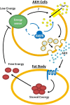
Model of AMPK function in AKH neuroendocrine cells. AMPK activation by low trehalose levels are detected through a currently unknown mechanism. Upon activation, AMPK leads to enhanced calcium levels in AKH cells, which subsequently leads to elevated levels of AKH release. The AKH hormone stimulates the fat body to release stored energy, which leads to the inhibition of AMPK and AKH hormone secretion.

References

    1. Alesutan I., Föller M., Sopjani M., Dërmaku-Sopjani M., Zelenak C., et al. , 2011. Inhibition of the heterotetrameric K+ channel KCNQ1/KCNE1 by the AMP-activated protein kinase. Mol. Membr. Biol. 28: 79–89 - PubMed
    1. Alzamora R., Gong F., Rondanino C., Lee J. K., Smolak C., et al. , 2010. AMP-activated protein kinase inhibits KCNQ1 channels through regulation of the ubiquitin ligase Nedd4–2 in renal epithelial cells. Am. J. Physiol. Renal Physiol. 299: F1308–F1319 - PMC - PubMed
    1. Andreelli F., Foretz M., Knauf C., Cani P. D., Perrin C., et al. , 2006. Liver adenosine monophosphate-activated kinase-alpha2 catalytic subunit is a key target for the control of hepatic glucose production by adiponectin and leptin but not insulin. Endocrinology 147: 2432–2441 - PubMed
    1. Bharucha K. N., Tarr P., Zipursky S. L., 2008. A glucagon-like endocrine pathway in Drosophila modulates both lipid and carbohydrate homeostasis. J. Exp. Biol. 211: 3103–3110 - PMC - PubMed
    1. Birk J. B., Wojtaszewski J. F., 2006. Predominant alpha2/beta2/gamma3 AMPK activation during exercise in human skeletal muscle. J. Physiol. 577: 1021–1032 - PMC - PubMed

Publication types

MeSH terms