. 2012 Oct;40(18):9329-39.
doi: 10.1093/nar/gks672. Epub 2012 Jul 14.

The solution structure of double helical arabino nucleic acids (ANA and 2'F-ANA): effect of arabinoses in duplex-hairpin interconversion

Affiliations

The solution structure of double helical arabino nucleic acids (ANA and 2'F-ANA): effect of arabinoses in duplex-hairpin interconversion

Nerea Martín-Pintado et al. Nucleic Acids Res. 2012 Oct.

Abstract

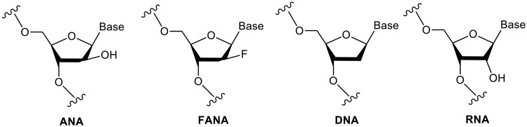
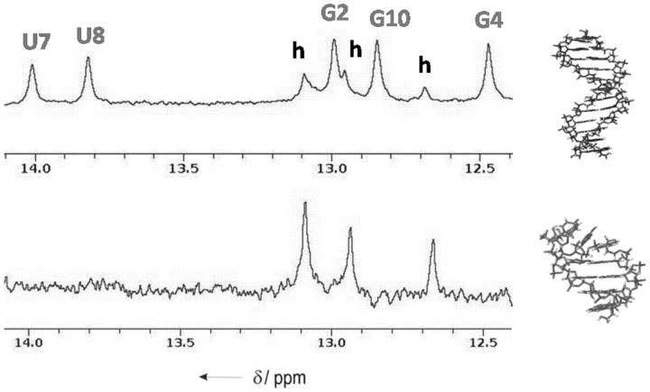
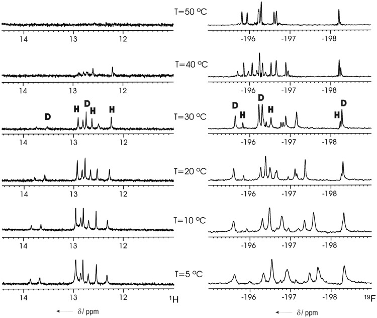
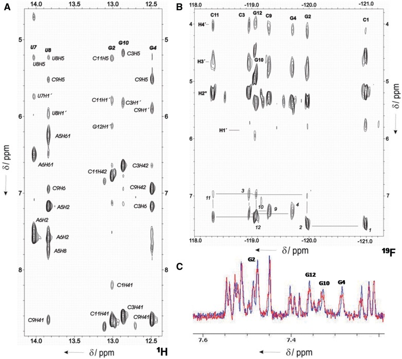
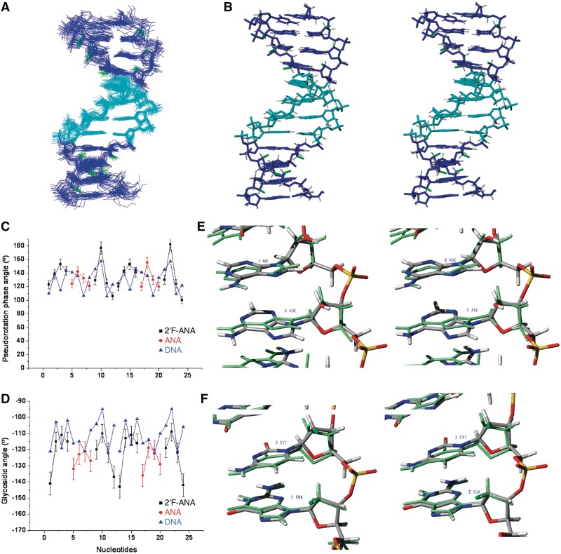
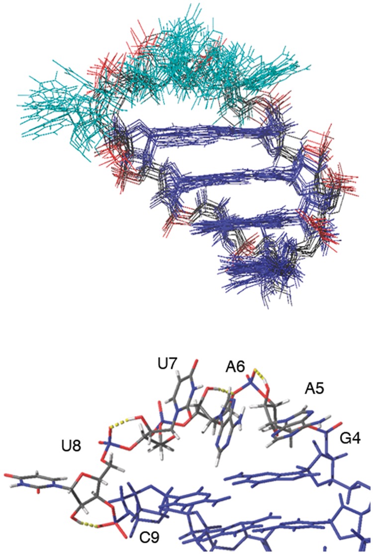
We report here the first structure of double helical arabino nucleic acid (ANA), the C2'-stereoisomer of RNA, and the 2'-fluoro-ANA analogue (2'F-ANA). A chimeric dodecamer based on the Dickerson sequence, containing a contiguous central segment of arabino nucleotides, flanked by two 2'-deoxy-2'F-ANA wings was studied. Our data show that this chimeric oligonucleotide can adopt two different structures of comparable thermal stabilities. One structure is a monomeric hairpin in which the stem is formed by base paired 2'F-ANA nucleotides and the loop by unpaired ANA nucleotides. The second structure is a bimolecular duplex, with all the nucleotides (2'F-ANA and ANA) forming Watson-Crick base pairs. The duplex structure is canonical B-form, with all arabinoses adopting a pure C2'-endo conformation. In the ANA:ANA segment, steric interactions involving the 2'-OH substituent provoke slight changes in the glycosidic angles and, therefore, in the ANA:ANA base pair geometry. These distortions are not present in the 2'F-ANA:2'F-ANA regions of the duplex, where the -OH substituent is replaced by a smaller fluorine atom. 2'F-ANA nucleotides adopt the C2'-endo sugar pucker and fit very well into the geometry of B-form duplex, allowing for favourable 2'F···H8 interactions. This interaction shares many features of pseudo-hydrogen bonds previously observed in 2'F-ANA:RNA hybrids and in single 2'F-ANA nucleotides.


Figures

Figure 1.
Figure 1.
Structures of ANA and 2′F-ANA in comparison with DNA and RNA.
Figure 2.
Figure 2.
Imino region of the 1H NMR spectra of 5′-fCfGfCfGaAaAaUaUfCfGfCfG-3′ at two different oligonucleotide concentrations: top, 0.9 mM and bottom, 0.2 mM (phosphate buffer 100 mM NaCl, pH = 7). Uppercase labels stands for duplex and lowercase labels for hairpin.
Figure 3.
Figure 3.
Imino region of the 1H (left) and complete 19F (right) NMR spectra of 5′-fCfGfCfGaAaAaUaUfCfGfCfG-3′ [gap(FA)] at different temperatures. Signals of duplex (D) and hairpin (H) species are indicated (oligonucleotide concentration of 0.8 mM, in the same buffer conditions as in Figure 2).
Figure 4.
Figure 4.
(A) Region of the NOESY spectrum of gap(FA) in H2O (mixing time: 150 ms). Watson–Crick base-pairs can be established for all the residues. (B) HOESY spectrum in D2O, showing the 19F-1H assignment pathway. (C) 19F-coupled (red) and 19F-decoupled (blue) 1H NMR spectra, demonstrating different signal intensity in H8 2′F-ANA purines. H8 of 2′F-ANA purines in the duplex are labelled. Oligonucleotide concentration of 0.8 mM, same buffer conditions as in Figure 2.
Figure 5.
Figure 5.
(A) Ensemble of the 20 refined structures of the duplex form of 5′-fCfGfCfGaAaAaUaUfCfGfCfG-3′. (B) Stereo view of the average structure. ANA nucleotides are shown in cyan, 2′F-ANA nucleotides in blue and fluorine atoms in green. (C) Pseudorotation phase angle and (D) glycosidic angle versus sequence. (E) Stereo view showing details of ANA residues, and (F) 2′F-ANA residues, superimposed to the unmodified DNA structure (in green colour, PDB: 1DUF).
Figure 6.
Figure 6.
Model of the hairpin structure of gap(FA). Top: ensemble of 10 structures from MD calculations. Bottom: detail of the ANA residues in the loop, showing hydrogen bonds between the 2′–OH and phosphate oxygens.

Similar articles

Cited by

References

    1. Noronha AM, Wilds CJ, Lok CN, et al. Synthesis and biophysical properties of arabinonucleic acids (ANA): circular dichroic spectra, melting temperatures, and ribonuclease H susceptibility of ANA·RNA hybrid duplexes. Biochemistry. 2000;39:7050–7062. - PubMed
    1. Watts JK, Damha MJ. 2′F-arabinonucleic acids (2′F-ANA)-history, properties, and new frontiers. Can. J. Chem. 2008;86:641–656.
    1. Neef AB, Luedtke NW. Dynamic metabolic labeling of DNA in vivo with arabinosyl nucleosides. Proc. Natl. Acad. Sci. USA. 2011;108:20404–20409. - PMC - PubMed
    1. Dowler T, Bergeron D, Tedeschi AL, et al. Improvements in siRNA properties mediated by 2′-deoxy-2′-fluoro-β-d-arabinonucleic acid (FANA) Nucleic Acids Res. 2006;34:1669–1675. - PMC - PubMed
    1. Kalota A, Karabon L, Swider CR, et al. 2′-deoxy-2′-fluoro-beta-D-arabinonucleic acid (2′F-ANA) modified oligonucleotides (ON) effect highly efficient, and persistent, gene silencing. Nucleic Acids Res. 2006;34:451–461. - PMC - PubMed

Publication types

Associated data