

Review
doi: 10.1136/bcr.07.2010.3155.
Longstanding rhinolith leading to anatomical alterations of the ipsilateral inferior nasal meatus and turbinate
Affiliations
- PMID: 22798515
- PMCID: PMC3029049
- DOI: 10.1136/bcr.07.2010.3155
Item in Clipboard
Review
Longstanding rhinolith leading to anatomical alterations of the ipsilateral inferior nasal meatus and turbinate
BMJ Case Rep.
.
Abstract
Rhinoliths consist of a central nidus with calcification resulting in calcareous concretions within the nasal cavity. They are uncommon in the literature despite a propensity particularly in children to insert foreign bodies into their nose and ears. We present the case of a 62-year-old woman with a longstanding undetected rhinolith with mild uncharacteristic symptoms. Radiographic examination revealed anatomical alteration of the inferior turbinate that was attributed to the long presence of the rhinolith in the nasal cavity. The management of the rhinolith and a review of the literature are presented.
Conflict of interest statement
Figures
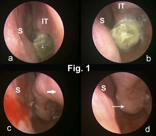
(A,B) Endoscopic view of the rhinolith on the floor of the left nasal cavity before and after mobilisation. (C) Left nasal cavity after removal of the rhinolith. Marked atrophy of the head (bold arrow) and anterior third of the inferior turbinate is visible. (D) Mild polypoid degeneration of the turbinate mucosa posterior to the rhinolith (slim arrow) can be seen. IT, inferior turbinate; S, septum.
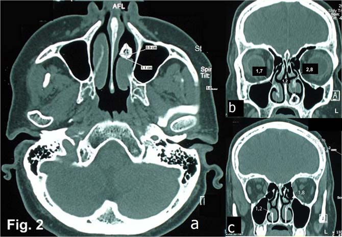
Axial CT image (A) shows a radiopaque object with a radiolucent core (anteroposterior, sagittal dimensions were 9×11 mm) in the anterior third of the left nasal cavity. Atrophy of mucosa and os turbinalis is visible. Coronal CT views of the anterior (B) and middle (C) third of the inferior nasal meatus show the height difference between the left (2.8 cm/1.8 cm) and right (1.7 cm/1.2 cm) meatus. No other anatomical alterations were detected.
Similar articles
-
[Wry nose and rhinolith: a case report].Lin Chuang Er Bi Yan Hou Tou Jing Wai Ke Za Zhi. 2017 Sep 5;31(17):1373-1375. doi: 10.13201/j.issn.1001-1781.2017.17.021. Lin Chuang Er Bi Yan Hou Tou Jing Wai Ke Za Zhi. 2017. PMID: 29798237 Chinese.
-
A bolt from the blew: rhinolith in the nose for more than 80 years.BMJ Case Rep. 2012 Nov 27;2012:bcr-2012-007322. doi: 10.1136/bcr-2012-007322. BMJ Case Rep. 2012. PMID: 23188861 Free PMC article.
-
Nasendoscopy for unusual nasal symptoms.BMJ Case Rep. 2010 Sep 9;2010:bcr0420102911. doi: 10.1136/bcr.04.2010.2911. BMJ Case Rep. 2010. PMID: 22778205 Free PMC article.
-
Rhinoliths causing palatal perforation: case report and literature review.Oral Surg Oral Med Oral Pathol Oral Radiol Endod. 2007 Dec;104(6):e42-6. doi: 10.1016/j.tripleo.2007.05.022. Epub 2007 Oct 17. Oral Surg Oral Med Oral Pathol Oral Radiol Endod. 2007. PMID: 17942340 Review.
-
Recurrent rhinolithiasis: a case report with review of the literature.West Indian Med J. 2012 Oct;61(7):760-3. West Indian Med J. 2012. PMID: 23620978 Review.
Cited by
-
Rock, paper, endoscopy: a baffling case of rhinolith.BMJ Case Rep. 2013 Apr 9;2013:bcr2013009147. doi: 10.1136/bcr-2013-009147. BMJ Case Rep. 2013. PMID: 23576662 Free PMC article.
-
Large rhinolith causing nasal obstruction.BMJ Case Rep. 2015 Mar 10;2015:bcr2014208260. doi: 10.1136/bcr-2014-208260. BMJ Case Rep. 2015. PMID: 25759270 Free PMC article.
-
Giant 'staghorn' rhinolith in a 15-year-old girl.BMJ Case Rep. 2018 Dec 14;11(1):e227587. doi: 10.1136/bcr-2018-227587. BMJ Case Rep. 2018. PMID: 30567272 Free PMC article.
-
A nose out of joint: first reported case of prison-acquired marijuana-based rhinolith.BMJ Case Rep. 2019 Oct 25;12(10):e231989. doi: 10.1136/bcr-2019-231989. BMJ Case Rep. 2019. PMID: 31653637 Free PMC article.
-
Rhinolith in the concha bullosa as a rare location: a case report.J Int Med Res. 2020 Aug;48(8):300060520951019. doi: 10.1177/0300060520951019. J Int Med Res. 2020. PMID: 32847434 Free PMC article.
References
-
- Hadi U, Ghossaini S, Zaytoun G. Rhinolithiasis: a forgotten entity. Otolaryngol Head Neck Surg 2002;126:48–51 - PubMed
-
- Shaw LC. Rhinolith of endogenous origin: A rare entity. Surg Pract 2007;11:48–50
-
- Yuca K, Caksen H, Etlik O, et al. The importance of rigid nasal endoscopy in the diagnosis and treatment of rhinolithiasis. Auris Nasus Larynx 2006;33:19–22 - PubMed
-
- Singh RK, Varshney S, Bist SS, et al. A case of rhinolithiasis. Online J Health Allied Scs 2008;7(2):7
Publication types
MeSH terms
LinkOut - more resources
Full Text Sources
Medical