

Review
doi: 10.1101/cshperspect.a008243.
Branching morphogenesis: from cells to organs and back
Affiliations
- PMID: 22798543
- PMCID: PMC3475165
- DOI: 10.1101/cshperspect.a008243
Item in Clipboard
Review
Branching morphogenesis: from cells to organs and back
Cold Spring Harb Perspect Biol.
.
Abstract
Many animal organs, such as the lung, the kidney, the mammary gland, and the vasculature, consist of branched tubular structures that arise through a process known as "branching morphogenesis" that results from the remodeling of epithelial or endothelial sheaths into multicellular tubular networks. In recent years, the combination of molecular biology, forward and reverse genetic approaches, and their complementation by live imaging has started to unravel rules and mechanisms controlling branching processes in animals. Common patterns of branch formation spanning diverse model systems are beginning to emerge that might reflect unifying principles of tubular organ formation.
Figures
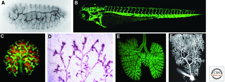
Branched animal organs. (A) Drosophila embryonic tracheal system stained with the tracheal luminal antigen mAb2A12, reproduced with permission from Development (Samakovlis et al. 1996a) (http://dev.biologists.org/content/122/5/1395 ). (B) Three-day-old zebrafish embryo vascular system expressing EGFP under the control of the endothelial specific promoter of the flk1 gene (TG:flk1:EGFP). (C) Developing mouse kidney stained for Wilm’s tumor 1 antigen (red, developing glomeruli) and calbindin (green, ureteric tree). (Photo courtesy of Kenneth Walker and John Bertram.) (D) Mammary gland of a virgin rat (Schedin et al. 2007). (From Schedin et al. 2007; adapted, with kind permission, from Springer Science+Business Media, J Mammary Gland Biol Neoplasia © 2007.) (E) Whole mount of an embryonic day 14.5 (E14.5) mouse lung stained for E-cadherin to show the airway epithelium. (Photo courtesy of Ross J. Metzger.) (F) Two-photon fluorescence image of Purkinje cell filled with a fluorescent dye. (Photo courtesy of Yo Otsu and Stéphane Dieudonné.)
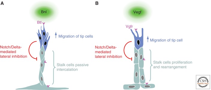
Branch regionalization and outgrowth. (A) Tracheal branching, and (B) vertebrate angiogenesis. Both Bnl and Vegf ligands elicit a migratory behavior through their respective receptors Btl and Vgfr. In both instances, the cells with the highest receptor activity become tip cells and produce more Delta, leading to the activation of Notch in neighboring stalk cells, which, in turn, inhibit the tip cell fate. Although Fgf signaling in the trachea triggers migration in the tip cells and, as a consequence, passive stalk cell intercalation, Vegf signaling in the vasculature not only triggers tip cell migration but also actively regulates the proliferation and rearrangement of stalk cells.
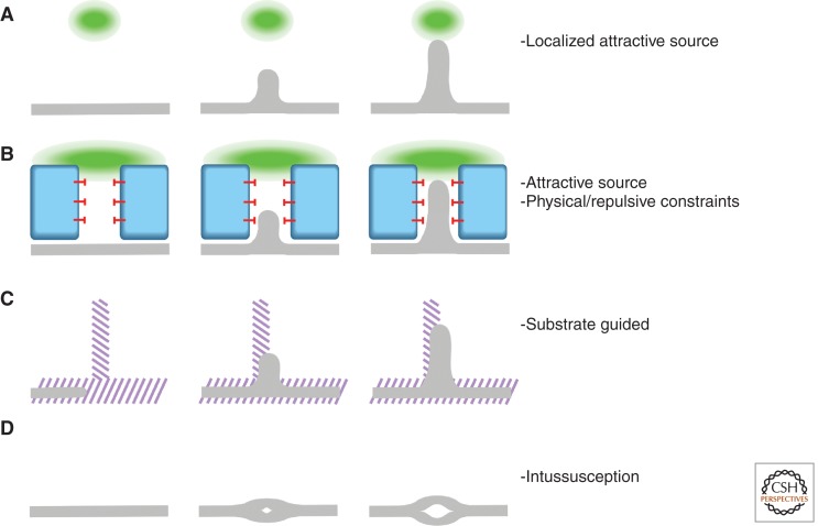
Branching strategies. Organ branching can be dissected into a few basic strategies. None of the branching events described in this review follow one single scheme but, rather, a combination of them. However, the identification within each organ of these prime processes can help us to understand and experimentally examine their development. (A) Migration toward a signaling source (green). (B) Migration toward a signaling source (green) constrained by repulsive signals (red block-arrows) or physical obstacles (blue). (C) Migration following an underlying patterned structure (purple stripes). (D) Intussusception: splitting of a branch into finer branches.
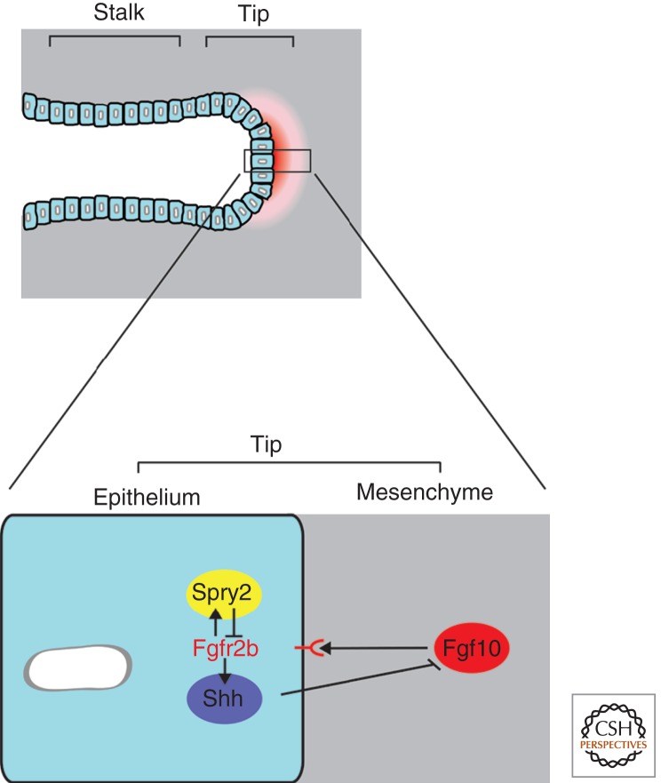
Molecular regulation of lung branching morphogenesis. A distal organizer and signaling center is located at the tip of the primary lung buds. At the core of this center is a set of reciprocal interactions between the bud epithelium and its surrounding mesenchyme. Fgf signaling promotes lung bud outgrowth; Fgf10 is expressed in the mesenchyme and signals to the epithelium through its receptor Fgfr2b. Fgf signaling increases Spry2 expression, which then antagonizes Fgf signaling in the epithelium. Additionally, Fgf signaling-induced Shh signals from the epithelium to the mesenchyme to negatively regulate Fgf10 expression in that tissue. Bmp4, on the other hand, seems to have a branching-promoting activity in the mesenchyme and an Fgf signaling down-regulation function within the epithelium, suggesting that correct Bmp4 levels are essential for lung development (not shown). The reciprocal interactions between a branching epithelium and the surrounding mesenchyme seem to be the organizing structural basis of many other budding organs such as the limb bud and the kidney.
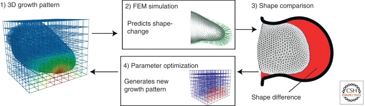
Limb development modeling through parameter exploration. Boehm et al. (2010) mapped theoretical proliferation values onto the observed initial limb shape (1). Using these values, the simulation produces a final limb shape that is compared with the observed final limb shape (2, 3); a parameter optimization step (4) generates a new initial proliferation pattern (1) that goes through the optimization process until a stable proliferation pattern is achieved. The simulations either fail or succeed to recapitulate the observed limb growth and the theoretical values, giving rise to a given shape that can be compared with the observed experimental data. (Reproduced from PLoS Biol., Boehm et al. 2010.)
References
-
- Affolter M, Caussinus E 2008. Tracheal branching morphogenesis in Drosophila: New insights into cell behaviour and organ architecture. Development 135: 2055–2064 - PubMed
-
- Affolter M, Mann R 2001. Development. Legs, eyes, or wings—selectors and signals make the difference. Science 292: 1080–1081 - PubMed
-
- Affolter M, Zeller R, Caussinus E 2009. Tissue remodelling through branching morphogenesis. Nat Rev Mol Cell Biol 10: 831–842 - PubMed
Publication types
MeSH terms
LinkOut - more resources
Full Text Sources
Molecular Biology Databases