. 2012 Jul 13;337(6091):236-9.
doi: 10.1126/science.1222981.

Molecular architecture and assembly principles of Vibrio cholerae biofilms

Affiliations

Molecular architecture and assembly principles of Vibrio cholerae biofilms

Veysel Berk et al. Science. .

Abstract

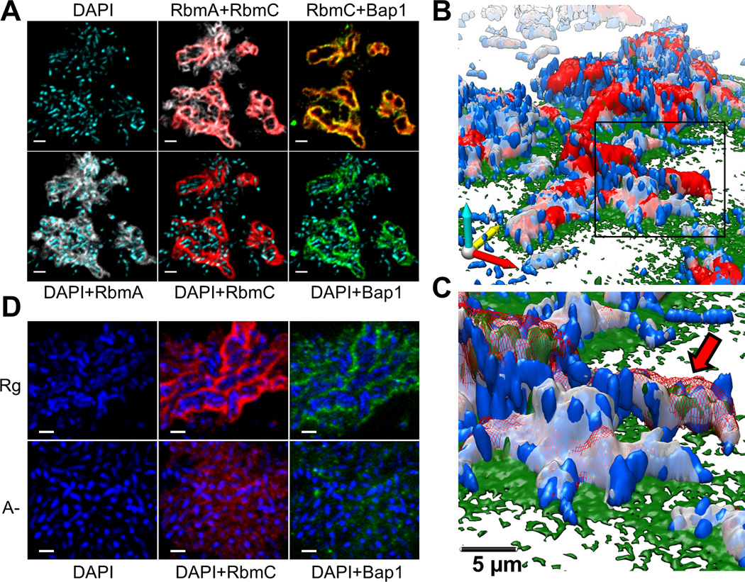
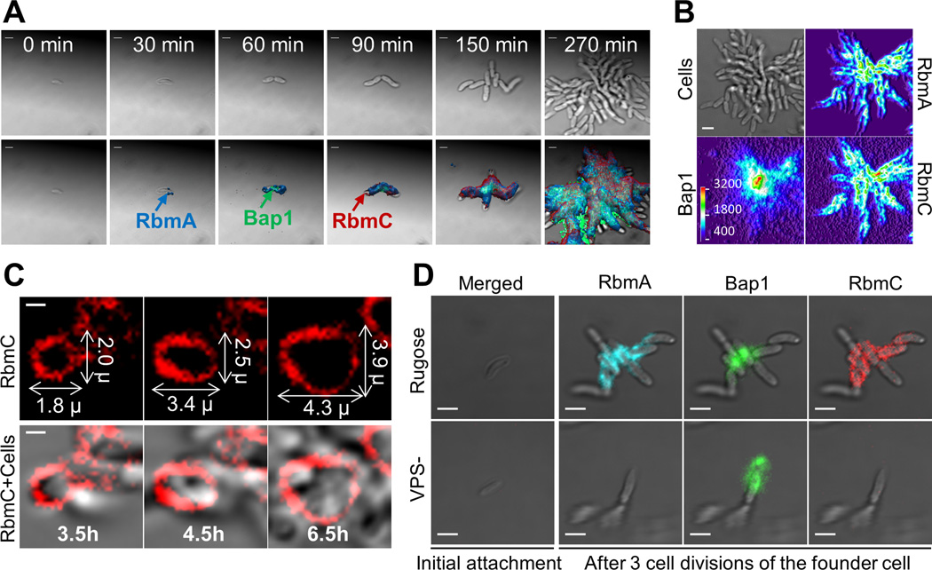
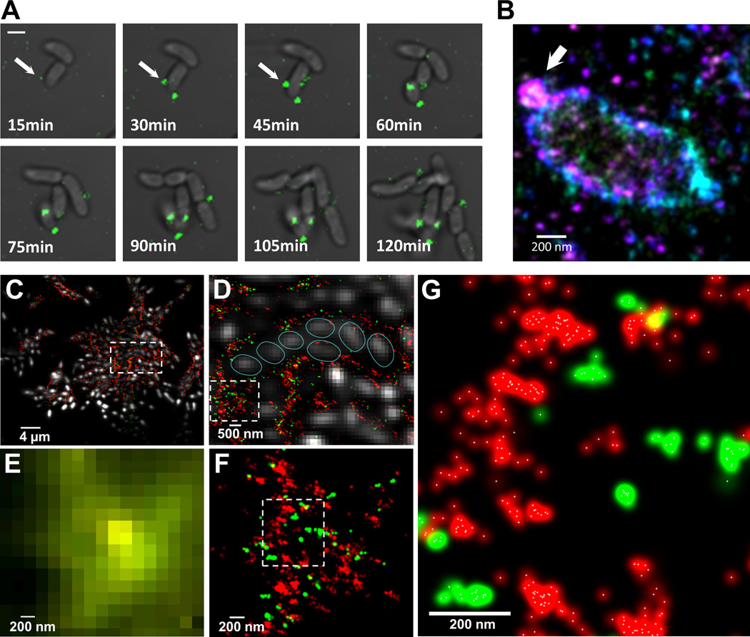
In their natural environment, microbes organize into communities held together by an extracellular matrix composed of polysaccharides and proteins. We developed an in vivo labeling strategy to allow the extracellular matrix of developing biofilms to be visualized with conventional and superresolution light microscopy. Vibrio cholerae biofilms displayed three distinct levels of spatial organization: cells, clusters of cells, and collections of clusters. Multiresolution imaging of living V. cholerae biofilms revealed the complementary architectural roles of the four essential matrix constituents: RbmA provided cell-cell adhesion; Bap1 allowed the developing biofilm to adhere to surfaces; and heterogeneous mixtures of Vibrio polysaccharide, RbmC, and Bap1 formed dynamic, flexible, and ordered envelopes that encased the cell clusters.


Figures

Fig. 1
Fig. 1. V. cholerae biofilm structure
(A) Optical section of biofilm 4 µm above coverslip. Images are pseudo-colored blue (cells), gray (RbmA), red (RbmC) and green (Bap1). RbmA localizes around and within cell clusters. RbmC and Bap1 encase cell clusters. Cells were counterstained with DAPI. Scale bars, 3 µm. (B) 3D biofilm architecture. Colors as in (B). (C) Enlargement of the boxed region in (B). Red arrow indicates one cell cluster. Red signal now rendered partially transparent to allow visualization of cells within an RbmC-containing cluster. (D) Comparison of biofilm architecture formed by rugose (Rg) and ΔrbmA (A-) strains. RbmA is required for cell cluster formation. Scale bars, 2 µm.
Fig. 2
Fig. 2. Time-lapse CLSM imaging of V. cholerae biofilm development and cluster formation
(A) Expression and subsequent distribution of matrix proteins were followed by time-lapse CLSM using continuous direct immunostaining. Cell outlines (bright field) are gray; RbmA, Bap1 and RbmC are shown in blue, green, and red, respectively. Scale bars, 2 µm. (B) Bright field biofilm image and corresponding fluorescent channel surface plots of Bap1, RbmA and RbmC obtained 4.5 hours post-inoculation. Fluorescent intensity is color-coded according to the color scale bar. Bap1 spread from a central point corresponding to the founder cell position while RbmA and RbmC were more homogenously distributed through the biofilm cells. Scale bar, 3 µm. (C) Gradual expansion of the RbmC-containing cluster tracked by time-lapse CLSM. Scale bars, 1 µm. (D) Inability to produce VPS (VPS-) prevents retention of daughter cells, accumulation of RbmA and RbmC, and blocks biofilm formation.
Fig. 3
Fig. 3. Exopolysaccharide secretion, initial organization and molecular architecture of V. cholerae biofilms
(A) Time-lapse CLSM images of VPS (green) production/secretion in V. cholerae cell during biofilm formation. Fluorescent images of VPS are merged with bright-field images of cells. Scale bars, 2 µm. (B) 3D superresolution image of a single V. cholerae cell. White arrow indicates a ball-like structure of VPS on the surface of V. cholerae cell early in biofilm formation. Color corresponds to height (−300 nm (violet) to +300nm (red)). (C) 3D two-color superresolution image (200 nm z-section) of a rugose variant biofilm showing molecular organization of VPS (red) and RbmC (green) around cell clusters. Cells were counterstained with DAPI (white). (D) Enlarged boxed region in (C) showing organization of cells within VPS/RbmC-enclosed cluster. Individual cells were outlined (cyan) for clarity. (E) Enlarged boxed region in (D) as it appears in conventional, diffraction-limited microcopy, showing unresolved VPS and RbmC signals. (F) Superresolution image of the same region in (E), showing distribution of RbmC and the VPS polymers in a biofilm matrix. (G) Enlarged boxed region in (F). White crosses indicate the center of a Gaussian-fit to each localization events.

Comment in

References

    1. Costerton JW, Stewart PS, Greenberg EP. Bacterial Biofilms: A Common Cause of Persistent Infections. Science. 1999;284:1318–1322. - PubMed
    1. Hall-Stoodley L, Stoodley P. Biofilm formation and dispersal and the transmission of human pathogens. Trends in Microbiology. 2005;13:7–10. - PubMed
    1. Parsek MR, Singh PK. Bacterial biofilms: An Emerging Link to Disease Pathogenesis. Annu. Rev. Microbiol. 2003;57:677–701. - PubMed
    1. Stewart PS, William Costerton J. Antibiotic resistance of bacteria in biofilms. The Lancet. 2001;358:135–138. - PubMed
    1. Mah T-F, et al. A genetic basis for Pseudomonas aeruginosa biofilm antibiotic resistance. Nature. 2003;426:306–310. - PubMed

Publication types

MeSH terms

Substances