. 2012 Oct 15;21(20):4378-93.
doi: 10.1093/hmg/dds271. Epub 2012 Jul 13.

Dystrophin and utrophin expression require sarcospan: loss of α7 integrin exacerbates a newly discovered muscle phenotype in sarcospan-null mice

Affiliations

Dystrophin and utrophin expression require sarcospan: loss of α7 integrin exacerbates a newly discovered muscle phenotype in sarcospan-null mice

Jamie L Marshall et al. Hum Mol Genet. .

Abstract

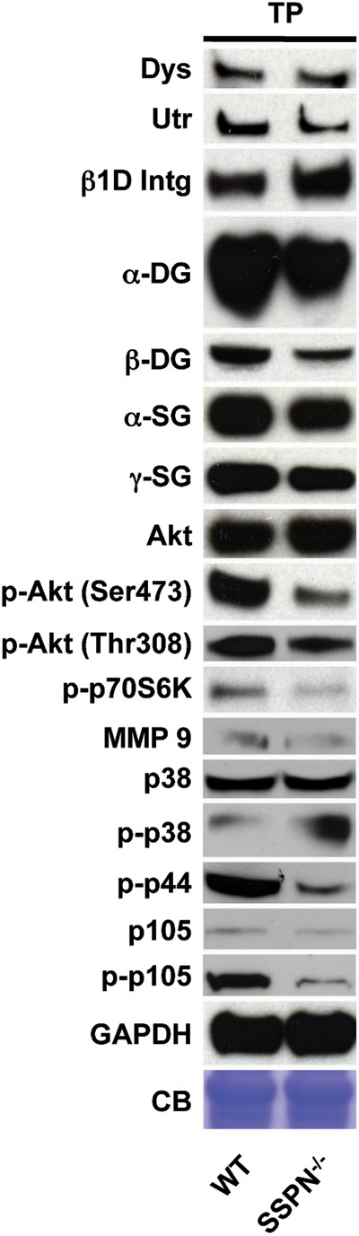
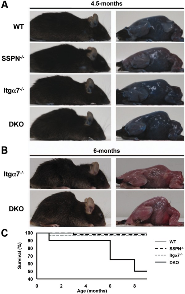
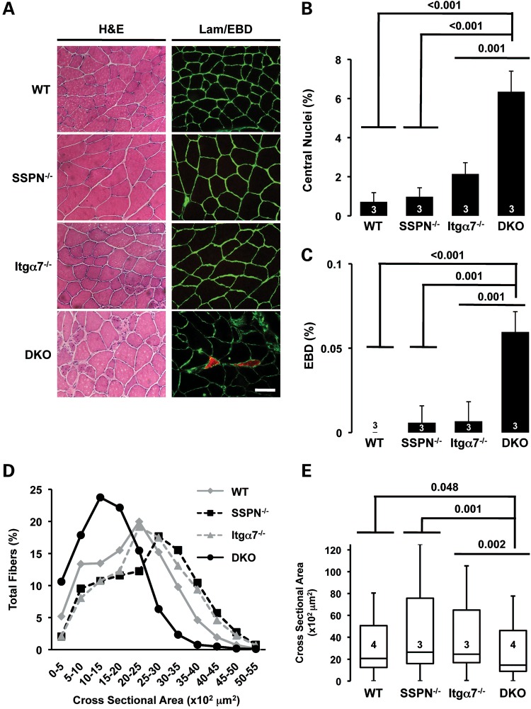
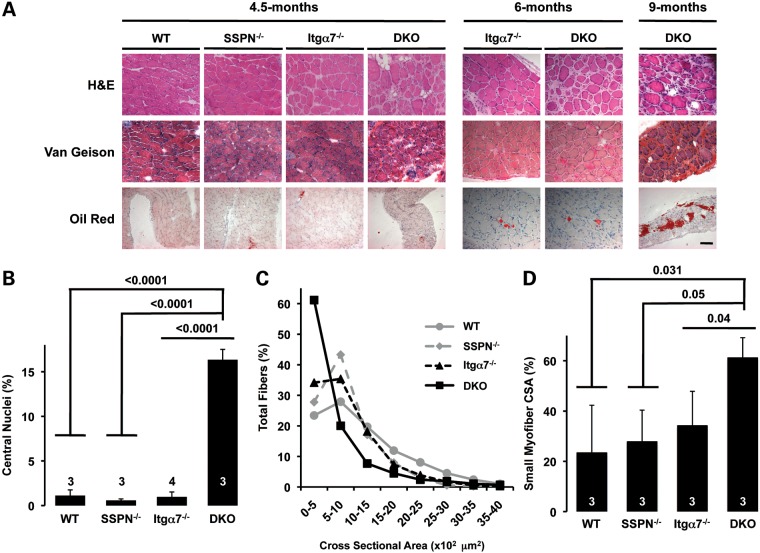
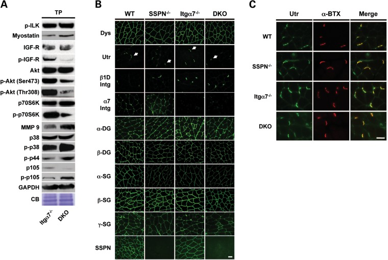
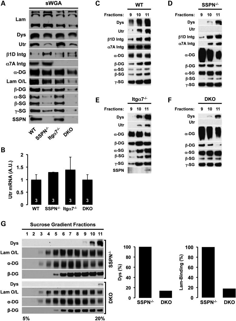
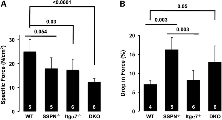
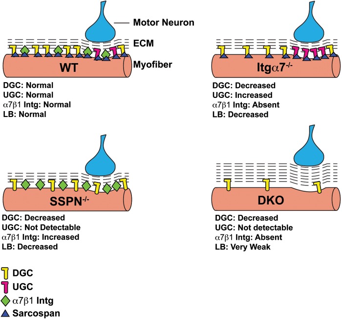
Sarcospan (SSPN) is a core component of the major adhesion complexes in skeletal muscle, the dystrophin- and utrophin (Utr)-glycoprotein complexes (DGC and UGC). We performed a rigorous analysis of SSPN-null mice and discovered that loss of SSPN decreased DGC and UGC abundance, leading to impaired laminin-binding activity and susceptibility to eccentric contraction-induced injury in skeletal muscle. We show that loss of SSPN increased levels of α7β1 integrin. To genetically test whether integrin compensates for the loss of DGC and UGC function in SSPN-nulls, we generated mice lacking both SSPN and α7 integrin (DKO, double knockout). Muscle regeneration, sarcolemma integrity and fibrosis were exacerbated in DKO mice and were remarkably similar to muscle from Duchenne muscular dystrophy (DMD) patients, suggesting that secondary loss of integrin contributes significantly to pathogenesis. Expression of the DGC and UGC, laminin binding and Akt signaling were negatively impacted in DKO muscle, resulting in severely diminished specific force properties. We demonstrate that SSPN is a necessary component of dystrophin and Utr function and that SSPN modulation of integrin signaling is required for extracellular matrix attachment and muscle force development.


Figures

Figure 1.
Figure 1.
Integrin is increased in SSPN-deficient muscle. Skeletal muscle lysates from 4.5-month-old wild-type (WT) and SSPN-null (SSPN−/−) mice were prepared in modified RIPA buffer. Equal protein samples (60 μg) were resolved by SDS–PAGE. Immunoblotting was performed with antibodies against dystrophin (Dys), utrophin (Utr), β1D integrin (β1D Intg), DGs (α- and β-DG), SGs (α- and γ-SG), Akt, phosphorylated Akt (p-Akt, Ser473), phosphorylated Akt (p-Akt, Thr308), phosphorylated p70S6K (p-p70S6K), matrix metalloproteinase 9 (MMP 9), p38 MAPK (p38), phosphorylated p38 MAPK (p-p38), phosphorylated p44 MAPK (p-p44), NF-κB1 p105 (p105), phosphorylated NF-κB1 p105 (p-p105). GAPDH and Coomassie blue (CB) staining serve as loading controls. Quantification of p-Akt/Akt, p-p70S6K and p-IGF-R/IGF-R are provided in Supplementary Material, Figure S1.
Figure 2.
Figure 2.
Severe kyphosis in SSPN- and α7 integrin-deficient (DKO) mice. (A) Photographs of wild-type (WT), SSPN-null (SSPN−/−), α7 integrin-null (Itgα7−/−) and SSPN-null:α7 integrin-null (DKO) mice at 4.5 months of age (left panels). Macroscopic evaluation of musculoskeletal structure in mice that were injected with EBD (right panels). DKO mice display severe kyphosis compared with control littermates. (B) Photographs of α7 integrin-null (Itgα7−/−) and SSPN-null:α7 integrin-null (DKO) mice at 6 months of age. DKO mice continue to exhibit severe kyphosis compared with control littermates. (C) The viability of DKO (n = 20) mice is severely reduced between 6 and 9 months of age compared with wild-type (n = 20), α7 integrin-null (n = 20) and SSPN-null (n = 20) mice. At 8 months, only 50% of DKO mice remain viable.
Figure 3.
Figure 3.
Enhanced muscle pathology in DKO mice compared with α7 integrin-null mice. (A) Transverse quadriceps sections from wild-type (WT), SSPN-null (SSPN−/−), α7 integrin-null (Itgα7−/−) and SSPN-null:α7 integrin-null (DKO) mice were stained for H&E to visualize nuclei and muscle pathology (left panels). Muscle cryosections from EBD-treated mice were incubated with laminin antibodies (green) to delineate the sarcolemma (right panels). EBD-positive fibers were visualized by red fluorescence, which serves as a marker for sarcolemma damage. Bar, 50 μm. (B) Muscle regeneration was evaluated by quantification of myofibers with central nuclei. Wild-type, SSPN-null and α7 integrin-null mice had <2% of fibers with centrally placed nuclei. The additional loss of SSPN resulted in a 3-fold increase in central nuclei in mice lacking α7 integrin (DKO). (C) Membrane damage was assayed by quantification of EBD uptake into quadriceps muscle fibers. Data are presented as an average and error bars represent standard deviation of the mean. (D) Myofiber CSA was quantified on transverse quadriceps cryosections stained with laminin to delineate myofiber boundaries for wild-type (WT) (n = 4), SSPN-null (SSPN−/−) (n = 3), α7 integrin-null (Itgα7−/−) (n = 3) and SSPN-null:α7 integrin-null (DKO) (n = 4) mice. SSPN-null and α7 integrin-null muscle exhibit myofiber hypertrophy compared with wild-type and DKO muscle. Average CSAs are plotted as an average percentage of total fibers (700 myofibers were analyzed per quadriceps). (E) Box plots demonstrating the variance and mean muscle fiber CSA of the data shown in (D). Median DKO myofibers are significantly hypotrophic compared with all controls. Boxes represent the middle quartiles from the 25th to 75th percentiles. Error bars represent maximum and minimum CSAs and statistics were calculated comparing the means. For (B)–(E), statistics were calculated using an ANOVA and Bonferroni's correction or Tukey's test. n- and P-values are indicated on the plots.
Figure 4.
Figure 4.
Increased fibrotic collagen deposition in the diaphragm of DKO mice. (A) Transverse cryosections of diaphragm muscle from wild-type (WT), SSPN-null (SSPN−/−), α7 integrin-null (Itgα7−/−) and SSPN-null:α7 integrin-null (DKO) were stained with H&E, Van Geison and Oil Red to visualize regeneration, collagen deposition and fat replacement, respectively. Age of the mice at the time of analysis is indicated. Bar, 50 μm. (B) Regeneration was quantified by counting central nuclei in 4.5-month-old diaphragms. Diaphragm muscles of DKO mice undergo significantly more regeneration (16-fold) compared with controls. (C) Myofiber CSAs of 4.5-month-old diaphragm muscles were quantified, revealing significant variation in fiber sizes in DKO muscles compared with controls. Small myofibers are prevalent in DKO (n = 3) diaphragm muscle compared with wild-type (n = 3), SSPN-null (n = 3) and α7 integrin-null (n = 3) controls. Average CSAs are plotted as an average percentage of total fibers (1400 myofibers were analyzed per diaphragm). (D) Graphs representing percentage of myofibers with very small CSAs (0–500 μm2). DKO diaphragms exhibit a 2-fold increase in very small myofibers compared with all controls. Error bars represent standard deviation of the mean. Statistics were calculated using an ANOVA with a Bonferroni correction or Tukey's test. n- and P-values are indicated on the plots.
Figure 5.
Figure 5.
Loss of SSPN and α7 integrin reduces the activation of Akt and levels of the DGC and UGC. (A) Skeletal muscle lysates from 4.5-month-old α7 integrin-null (Itgα7−/−) and SSPN-null:α7 integrin-null (DKO) mice were prepared in modified RIPA buffer. Equal protein samples (60 μg) were resolved by SDS–PAGE. Immunoblotting was performed with antibodies against phosphorylated ILK (p-ILK), myostatin, insulin-like growth factor receptor (IGF-R), phosphorylated IGF-R (p-IGF-R), Akt, phosphorylated Akt (p-Akt, Ser473), phosphorylated Akt (p-Akt, Thr308), p70S6K, phosphorylated p70S6K (p-p70S6K), MMP 9, p38 MAPK (p38), phosphorylated p38 MAPK (p-p38), phosphorylated p44 MAPK (p-p44), NF-κB1 p105 (p105) and phosphorylated NF-κB1 p105 (p-p105). Coomassie blue (CB) staining and GAPDH were used for loading controls. Quantification of p-Akt/Akt, p-p70S6K and p-IGF-R/IGF-R is provided in Supplementary Material, Figure S1. (B) Transverse cryosections of quadriceps muscle from 4.5-month-old wild-type (WT), SSPN-null (SSPN−/−), α7 integrin-null (Itgα7−/−) and SSPN-null:α7 integrin-null (DKO) mice were stained with antibodies against dystrophin (Dys), Utr, β1D integrin (β1D Intg), α7 integrin (α7 Intg), DGs (α- and β-DG), SGs (α-, β- and γ-SG) and SSPN. Arrows denote NMJ structures. Bar, 50 μm. (C) Transverse cryosections of quadriceps muscle from 4.5-month-old mice were co-stained with Utr antibody and α-BTX, which serve as a marker for NMJs. Merged images (right panels) reveal that Utr protein is localized to NMJ structures in all samples. However, NMJs in DKO muscle appear to be smaller in size with faint Utr staining. Bar, 50 μm.
Figure 6.
Figure 6.
SSPN and integrin stabilize protein interactions within the DGC and UGC. (A) Skeletal muscle lysates from 4.5-month-old wild-type (WT), SSPN-null (SSPN−/−), α7 integrin-null (Itgα7−/−) and SSPN-null:α7 integrin-null (DKO) mice were prepared in digitonin buffer and enriched by sWGA lectin chromatography. Equal protein samples (10 μg) were resolved by SDS–PAGE. Immunoblotting was performed with antibodies against laminin (Lam), dystrophin (Dys), Utr, α7 integrin (α7 Intg), β1D integrin (β1D Intg), DGs (α- and β-DG), SGs (α-, β-, γ-SG) and SSPN. Laminin protein was overlaid on α-DG and visualized by immunoblotting with antibody against laminin (Lam O/L). Quantification of lectin-purified adhesion complexes is provided in Supplementary Material, Figure S5. (B) Utr mRNA was measured from the quadriceps muscle of 4.5-month-old wild-type (WT), SSPN-null (SSPN−/−), α7 integrin-null (Itgα7−/−) and SSPN-null:α7 integrin-null (DKO) mice and normalized to the GAPDH internal control. Data are presented as an average normalized to wild-type, and error bars represent standard deviation of the mean (n = 3 mice per genotype). Levels of Utr mRNA were not statistically altered in any genotype. sWGA lectin enrichments from wild-type (C), SSPN-null (D), α7 integrin-null (Itgα7−/−) (E) and DKO (F) muscle were subjected to 5–20% sucrose gradient ultracentrifugation to investigate the integrity of glycoprotein adhesion complexes. Nitrocellulose transfers were probed with the indicated antibodies. Fraction numbers from the sucrose gradients are listed above the blots. The exposures shown are not identical between different genotypes. (G) Sucrose gradient fractions of sWGA eluates are displayed for SSPN-null and DKO mice. All blotting and exposures were performed under identical conditions to facilitate the comparison of relative intensities for the migration profiles of dystrophin (Dys), laminin binding to α-DG (Lam O/L) and DGs (α- and β-DG). Densitometry was performed on dystrophin-containing fractions for dystrophin and laminin binding. Data are represented as total percent band intensity relative to SSPN-null band intensity. DKO mice demonstrate an 87% reduction in dystrophin protein and an 83% reduction in laminin binding to α-DG compared with SSPN-deficient controls, as shown in the plots.
Figure 7.
Figure 7.
Specific force production is diminished in all mutant mice. (A) Specific force generation was measured from diaphragm muscles of 4.5-month-old wild-type (WT), SSPN-null (SSPN−/−), α7 integrin-null (Itgα7−/−) and SSPN-null:α7 integrin-null (DKO) mice. There is a significant reduction in the generation of specific force from α7 integrin-null and DKO mice compared with wild-type controls. Data represent averages and error bars represent standard deviation of the mean. Statistics were calculated using an ANOVA with a Bonferroni correction. n- and P-values are indicated. (B) The percentage drop in force between the first and fifth eccentric contraction in isolated diaphragm muscles of 4.5-month-old wild-type (WT), SSPN-null (SSPN−/−), α7 integrin-null (Itgα7−/−) and SSPN-null:α7 integrin-null (DKO) mice is plotted. The drop in force of SSPN-deficient and DKO diaphragms was significant compared with wild-type diaphragms. Data are presented as an average, and error bars represent standard deviation of the mean. Statistics were calculated using an ANOVA with a Bonferroni correction. n- and P-values are indicated on the plots.
Figure 8.
Figure 8.
Effect of SSPN and α7β1 integrin on adhesion complexes in muscle. A schematic diagram is provided to illustrate the adhesion glycoprotein complexes at the sarcolemma of 4.5-month-old wild-type (WT), SSPN-null (SSPN−/−), α7 integrin-null (Itgα7−/−) and SSPN-null:α7 integrin-null (DKO) mice. Protein levels of the entire DGC (yellow), entire UGC (pink) and α7β1 integrin (green) complexes are described relative to wild-type muscle. The presence of SSPN is indicated with dark-blue triangles. The dashed lines represent the amount of ECM or fibrosis, and the levels of laminin binding (LB) to α-DG are described. Extra-synaptic and NMJ (where motor neuron innervates muscle) regions of the sarcolemma are depicted. The levels of the UGC and DGC are reduced in SSPN-deficient and DKO mice, and α7β1 integrin is increased in SSPN-null muscle. In DKO muscle, the increased number of dashed lines represents the increase in fibrosis observed in the diaphragm muscle. Laminin binding to α-DG is weakest in DKO muscle due to the severe reductions observed in the levels of the DGC and UGC.

Similar articles

Cited by

References

    1. Bonnemann C.G., Finkel R.S. Sarcolemmal proteins and the spectrum of limb-girdle muscular dystrophies. Semin. Pediatr. Neurol. 2002;9:81–99. doi:10.1053/spen.2002.33795. - DOI - PubMed
    1. Carmignac V., Durbeej M. Cell-matrix interactions in muscle disease. J. Pathol. 2012;226:200–218. doi:10.1002/path.3020. - DOI - PubMed
    1. Ervasti J.M. Dystrophin, its interactions with other proteins, and implications for muscular dystrophy. Biochem. Biophys. Acta. 2007;1772:108–117. - PubMed
    1. Ibraghimov-Beskrovnaya O., Ervasti J.M., Leveille C.J., Slaughter C.A., Sernett S.W., Campbell K.P. Primary structure of dystrophin-associated glycoproteins linking dystrophin to the extracellular matrix. Nature. 1992;355:696–702. doi:10.1038/355696a0. - DOI - PubMed
    1. Ibraghimov-Beskrovnaya O., Milatovich A., Ozcelik T., Yang B., Koepnick K., Francke U., Campbell K.P. Human dystroglycan: skeletal muscle cDNA, genomic structure, origin of tissue specific isoforms and chromosomal localization. Hum. Mol. Genet. 1993;2:1651–1657. doi:10.1093/hmg/2.10.1651. - DOI - PubMed

Publication types

MeSH terms