. 2012 Oct 15;21(20):4406-18.
doi: 10.1093/hmg/dds273. Epub 2012 Jul 13.

Proteomic, genomic and translational approaches identify CRMP1 for a role in schizophrenia and its underlying traits

Affiliations

Proteomic, genomic and translational approaches identify CRMP1 for a role in schizophrenia and its underlying traits

Verian Bader et al. Hum Mol Genet. .

Abstract

Schizophrenia is a chronic illness of heterogenous biological origin. We hypothesized that, similar to chronic progressive brain conditions, persistent functional disturbances of neurons would result in disturbed proteostasis in the brains of schizophrenia patients, leading to increased abundance of specific misfolded, insoluble proteins. Identification of such proteins would facilitate the elucidation of molecular processes underlying these devastating conditions. We therefore generated antibodies against pooled insoluble proteome of post-mortem brains from schizophrenia patients in order to identify unique, disease-specific epitopes. We successfully identified such an epitope to be present on collapsin-response mediator protein 1 (CRMP1) in biochemically purified, insoluble brain fractions. A genetic association analysis for the CRMP1 gene in a large Finnish population cohort (n = 4651) corroborated the association of physical and social anhedonia with the CRMP1 locus in a DISC1 (Disrupted-in-schizophrenia 1)-dependent manner. Physical and social anhedonia are heritable traits, present as chronic, negative symptoms of schizophrenia and severe major depression, thus constituting serious vulnerability factors for mental disease. Strikingly, lymphoblastoid cell lines derived from schizophrenia patients mirrored aberrant CRMP1 immunoreactivity by showing an increase of CRMP1 expression, suggesting its potential role as a blood-based diagnostic marker. CRMP1 is a novel candidate protein for schizophrenia traits at the intersection of the reelin and DISC1 pathways that directly and functionally interacts with DISC1. We demonstrate the impact of an interdisciplinary approach where the identification of a disease-associated epitope in post-mortem brains, powered by a genetic association study, is rapidly translated into a potential blood-based diagnostic marker.


Figures

Figure 1.
Figure 1.
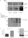
(A) Dot blot of a pool of normal (n = 15; left) and schizophrenia-diagnosed (n = 15; right) brain homogenates, incubated with different hybridoma supernatants containing monoclonal antibodies (as examples: #A–#F). Selection of one partial dot blot from a total of approximately 1300 different clones screened. Although some antibodies recognized both normal and schizophrenia-diagnosed brain homogenates equally weak (#B, #C, #E, #F), one recognized both brain homogenate pools strongly but without distinction (#D). Clone #A recognized the schizophrenia homogenate pool much better than the normal control. This mAB was subsequently taken forward for epitope identification (see also Supplementary Material, Table S1 and Fig. S1). (B) Western blots of recombinant GST-CRMP1 recombinantly expressed and purified from E. coli and probed with mAB #A, α-CRMP1 mAB 4C12 (generated in-house) and commercial rabbit α-CRMP1 antibody (ProSci #3625) as indicated. (C) Western blot of mouse brain homogenate in which a lane was cut in half, with the left part being incubated with a commercial rabbit α-CRMP1 antibody (ProSci #3625) and the right part incubated with mAB #A. The blot shows that immunoreactivity at the same molecular weight is stained by both antibodies, corroborating the identity of the mAB #A antigen as being CRMP1. (D) Western blot of insoluble fractions (‘pellet’) or starting material homogenate (‘lysate’) from 60 post-mortem brains with schizophrenia (S), bipolar disorder (B), depression (D) or normal controls (N); compare with (E). Antibody: rabbit α-CRMP1 (ProSci #3625). (E) Quantitative immunoreactivity of α-CRMP1 antibody against purified, insoluble protein pellets from equal starting material of post-mortem brains from the SMRI CC for chronic mental illness patients (n = 45) and normal controls (n = 15). Densitometry was performed from original western blots (D). Resulting measurements were normalized against the expression level of CRMP1 in the starting material, i.e. the brain homogenate (‘lysate’) (B). Expression in diseased samples reached statistical significance with P < 0.05 (ranked Mann–Whitney one-tailed since samples were not equally distributed).
Figure 2.
Figure 2.
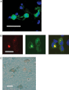
(A) Western blot of neuroblastoma cell fractions (total lysate, supernatant and insoluble pellet) transiently transfected with untagged CRMP1sv alone (lanes 1, 4 and 7), untagged DISC1 alone (lanes 2, 5 and 8) or both untagged CRMP1sv and DISC1 (lanes 3, 6 and 9). The blot shows that although CRMP1sv alone has no tendency to form insoluble pellets (lane 7), in the presence of insoluble DISC1, CRMP1sv is recruited into the purified insoluble pellet, demonstrating an interaction between insoluble DISC1 and CRMP1sv (lane 9). (B) Western blot of a pulldown experiment performed with a recombinant His6-tagged, human DISC1 fragment (amino acids 316–854) immobilized on NHS Sepharose (or non-DISC1-linked NHS as a negative control, ‘NHS blocked’) and incubated with either GST alone (‘GST’) or fusion protein GST-CRMP1 (‘GST-CRMP1’). Immobilized His6-tagged DISC1(316–854) specifically pulls down GST-CRMP1, demonstrating a physical interaction in this cell-free system. (C) Compilation of four independent analyses of primary mouse neurons silenced for DISC1 (see western blot in the right panel; antibody rabbit anti-mouse DISC1 antiserum #440 (76) for 72 h, measuring CRMP1 staining intensity. CRMP1 expression is clearly increased after DISC1 silencing. Mann–Whitney, two-tailed, P = 0.03.
Figure 3.
Figure 3.
(A) Fluorescent image of CAD cells transiently transfected with eGFP-CRMP1sv, demonstrating its solubility by its regular distribution within the cell. Bar, 20 μm. (B) Fluorescent image of CAD cells co-transfected with fusion proteins of eGFP-CRMP1sv (green) and mRFP-DISC1 (red). When co-expressed with mRFP-DISC1, eGFP-CRMP1sv is detected in aggresomes (34) and is not regularly dispersed throughout the cell as in Fig. 2A, demonstrating recruitment of eGFP-CRMP1 into mRFP-DISC1 aggresomes (merge to yellow, rightmost panel). (C) Immunohistochemistry with α-CRMP1 (green) and α-DISC1 (brown) of a human brain demonstrating co-localization of CRMP1 and DISC1 in cortical neurons. α-DISC1 mAB 3D4 (31) and rabbit α-CRMP1 (#3625, ProSci, USA) were used with DAB and HistoGreen as substrates, respectively. Bar, 20 μm.
Figure 4.
Figure 4.
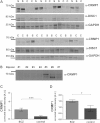
(A) Representative western blot of whole lymphoblastoid cell lysates from patients with schizophrenia (S) or normal controls (C) probed with α-CRMP1 antibody (#3625, ProSci), α-DISC1 antibody 14F2 (34) and α-GAPDH antibody as loading control (lower panel), as indicated. (B) Western blot of whole lymphoblastoid lysates from patients with bipolar disorder probed with α-CRMP1 antibody (#3625, ProSci). The western blot identifies cases #5 and #6 as positive for CRMP1 immunoreactivity. (C) Quantitative densitometry of α-CRMP1 immunoreactivity of lymphoblasts from schizophrenic patients (n = 14) and normal controls (n = 15), normalized by immunoreactivity to GAPDH [samples comprise those in (A)]. Lymphoblasts from patients with schizophrenia displayed significantly higher CRMP1 immunoreactivity. ***Mann–Whitney U test (no Gaussian distribution), two-tailed P < 0.001. (D) Quantitative densitometry of α-CRMP1 (#3625, ProSci) of an entirely independent collection of lymphoblastoid cell lines (University of Düsseldorf, Germany) from those in (A) and (C) (Johns Hopkins Medical School, USA), demonstrating significant, disease-associated differences in CRMP1 immunoreactivity (see Supplementary Material, Table S6 for demographic information).
Figure 5.
Figure 5.
Schematic drawing of molecular interactions between the DISC1 and reelin pathways. Direct functional connections in solid arrows, indirect functional connections (over several or unknown steps) in broken line arrows. The proteins depicted are an incomplete selection and focus on the so-far best described interactions.

References

    1. Freimer N., Sabatti C. The human phenome project. Nat. Genet. 2003;34:15–21. - PubMed
    1. Gottesman I.I., Gould T.D. The endophenotype concept in psychiatry: etymology and strategic intentions. Am. J. Psychiatry. 2003;160:636–645. - PubMed
    1. Fanous A., Gardner C., Walsh D., Kendler K.S. Relationship between positive and negative symptoms of schizophrenia and schizotypal symptoms in nonpsychotic relatives. Arch. Gen. Psychiatry. 2001;58:669–673. - PubMed
    1. Vollema M.G., Sitskoorn M.M., Appels M.C., Kahn R.S. Does the Schizotypal Personality Questionnaire reflect the biological-genetic vulnerability to schizophrenia? Schizophr. Res. 2002;54:39–45. - PubMed
    1. Kendler K.S., McGuire M., Gruenberg A.M., O'Hare A., Spellman M., Walsh D. The Roscommon Family Study. I. Methods, diagnosis of probands, and risk of schizophrenia in relatives. Arch. Gen. Psychiatry. 1993;50:527–540. - PubMed

Publication types