. 2012 Jul 11:6:24.
doi: 10.3389/fnana.2012.00024. eCollection 2012.

Excitatory neuronal connectivity in the barrel cortex

Affiliations

Excitatory neuronal connectivity in the barrel cortex

Dirk Feldmeyer. Front Neuroanat. .

Abstract

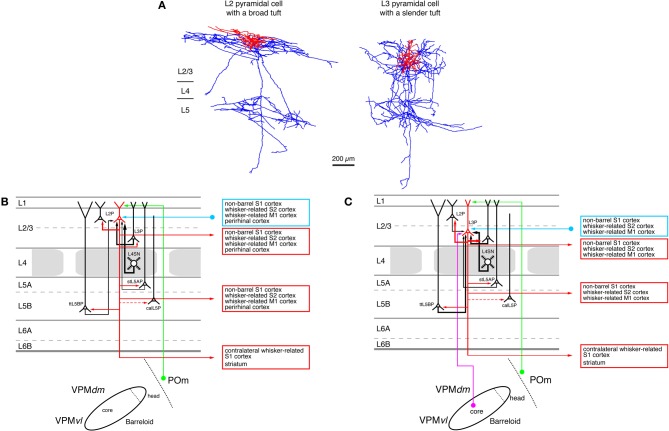
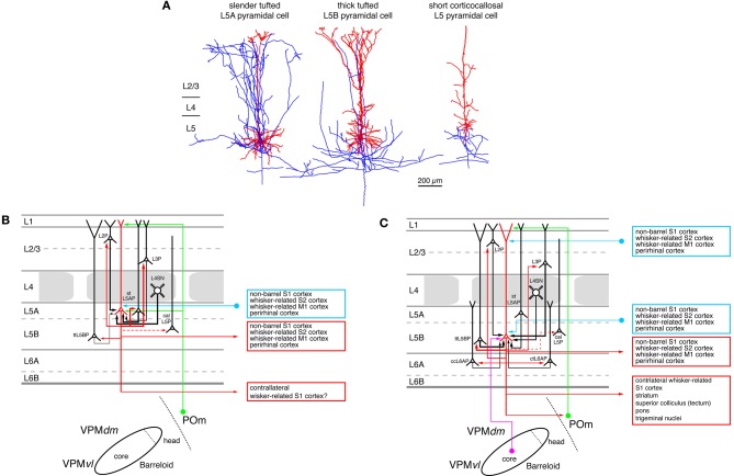
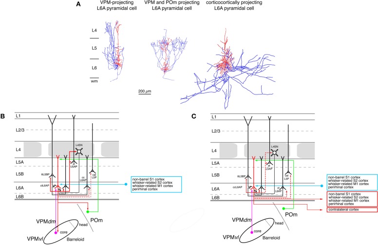
Neocortical areas are believed to be organized into vertical modules, the cortical columns, and the horizontal layers 1-6. In the somatosensory barrel cortex these columns are defined by the readily discernible barrel structure in layer 4. Information processing in the neocortex occurs along vertical and horizontal axes, thereby linking individual barrel-related columns via axons running through the different cortical layers of the barrel cortex. Long-range signaling occurs within the neocortical layers but also through axons projecting through the white matter to other neocortical areas and subcortical brain regions. Because of the ease of identification of barrel-related columns, the rodent barrel cortex has become a prototypical system to study the interactions between different neuronal connections within a sensory cortical area and between this area and other cortical as well subcortical regions. Such interactions will be discussed specifically for the feed-forward and feedback loops between the somatosensory and the somatomotor cortices as well as the different thalamic nuclei. In addition, recent advances concerning the morphological characteristics of excitatory neurons and their impact on the synaptic connectivity patterns and signaling properties of neuronal microcircuits in the whisker-related somatosensory cortex will be reviewed. In this context, their relationship between the structural properties of barrel-related columns and their function as a module in vertical synaptic signaling in the whisker-related cortical areas will be discussed.

Keywords: barrel cortex; cortical column; excitatory connections; long-range collaterals; pyramidal cell; somatosensory cortex; spiny stellate cell.


Figures

Figure 1
Figure 1
Dual labeling of axons projecting from the VPM and POm axons. Labeling of VPM and POm axons in the same animal by adeno-associated virus-mediated expression of different fluorescent proteins. VPM afferents (red) in a thalamocortical barrel cortex slice. POm afferents (green). Overlay of VPM and POm labeled thalamocortical axons illustrating afferent sparse zones of low fluorescence (i.e., low thalamocortical innervation). There is potential overlay of VPM and POm afferents in the deeper portion of barrels (yellow). Modified from Wimmer et al. (2010), with permission of the Society for Neuroscience.
Figure 2
Figure 2
Pathways from thalamus to the primary somatosensory cortex (S1). The figure shows the section of the four different whisker-related pathways from the thalamus to the primary somatosensory cortex. The thalamus is represented by a single barreloid in VPM; the border between POm and VPM by a dashed line. The input stations in the brainstem nuclei have been omitted in this diagram. Magenta: lemniscal (1) pathway; orange: lemniscal (2) pathway; green: paralemniscal pathway; blue: extralemniscal pathway. The term “dysgranular cortex” in S1 defines the region in and around the barrel field in which layer 4 shows no clear barrel structure. Abbreviations: VPMvl, ventrolateral portion of a barreloid in the ventroposterior medial nucleus of the thalamus; VPMdm, dorsomedial portion of a barreloid in the VPM.
Figure 3
Figure 3
Neuronal elements in the S1 barrel cortex with a predominantly vertical axonal organization. The figure shows four types of axonal projections (blue) with a predominantly vertical axonal projection that is largely confined to a barrel column in the whisker-related S1 cortex. (A) Most thalamic afferents from VPM nucleus of the thalamus arborize extensively in layer 4 in a barrel-restricted fashion, (B) L4 spiny stellate cell, (C) L4 star pyramidal cell, (D) corticothalamically projecting L6A pyramidal cell. The dendritic domain of intracortical neurons is given in red. (Modified from Oberlaender et al. (2011b) (A), from Feldmeyer et al. (1999) (B,C), from Zhang and Deschênes (D) with permission of John Wiley and sons on behalf of The Physiological Society, the Society for Neuroscience and Oxford Journals).
Figure 4
Figure 4
Excitatory synaptic input–output relationship in layer 4 of the S1 barrel cortex. (A) Reconstructions of a L4 spiny stellate cell (left) and a L4 star pyramidal neuron (right) in rat barrel cortex (Feldmeyer et al., 1999). Modified with permission of John Wiley and Sons on behalf of The Physiological Society. (B) Diagram of the excitatory synaptic connections of and onto L4 spiny neurons (red neuron with blue axon) in the barrel cortex. Although layer 4 contains both spiny stellate and star pyramidal neurons and a few pyramidal cells only spiny stellate cells are shown for simplicity. Note that L4 spiny neurons provide synaptic output to virtually all layers in a barrel column. For detailed information on the location of synaptic contacts and differences in the connectivity of the three different excitatory L4 neurons see text. The thalamic region is represented by a single barreloid in the VPM nucleus of the thalamus; the VPM/POm border is marked by a dashed line. Red neuron; Dendrites and axon of the neuron for which the input–output relationship is described in this figure. Different cortical layers as indicated on the left. The thickness of the red arrows pointing to a postsynaptic (black) neurons indicates the connection probability between this and the black neurons as well as cortical and subcortical areas. The dashed red arrow in layer 5 marks a likely but not yet verified synaptic connection onto a corticocallosal L5 pyramidal cell. It should be noted that Black neurons: Dendrites and axon of neurons sending to and receiving synaptic input from to the red neuron. The thickness of the black arrows pointing to the red neuron indicates the connection probability between these neurons and the red neuron. Light blue arrows: Excitatory synaptic input from cortical regions outside the S1 barrel cortex. Magenta arrow: Synaptic input from the VPM (lemniscal (1) pathway. Green arrow: Synaptic input from the POm (paralemniscal pathway). However, for L4 spiny neurons synaptic input from outside thhe barrel cortex originates almost exclusively from the core of the barreloid in the dorsomedial part of the VPM. Abbreviations: VPM, ventroposterior medial nucleus of the thalamus; dm, dorsomedial part; vl, ventrolateral part; POm, posterior medial nucleus of the thalamus; L2P, L2 pyramidal cell; L3P, L3 pyramidal cell; L4SN, L4 spiny neuron; stL5P, slender-tufted L5A pyramidal cell; ttL5BP, thick-tufted L5B pyramidal cell; calL5P, corticocallosal L5 pyramidal cell; ccL6AP, corticocortical L6A pyramidal cell; ctL6AP, corticothalamic L6A pyramidal cell.
Figure 5
Figure 5
Excitatory synaptic input–output relationship in layer 2/3 of the S1 barrel cortex. (A) Reconstructions of a pyramidal cell located in the upper half of layer 2/3 (L2 pyramidal cell, left) and a pyramidal cell located in the lower half of layer 2/3 (L3 pyramidal cell, left) of rat barrel cortex (Bruno et al., 2009); modified with permission of the Society of Neuroscience. Note that the apical tuft of the L2 pyramidal cell is substantially larger than the basal dendritic tree of that neuron while L3 pyramidal cells have slender apical tufts. Modified with permission from the Society of Neuroscience. (B) Diagram of the excitatory synaptic connections of and onto L2 pyramidal cells (red neuron with blue axon) in the barrel cortex. Only synaptic input from neurons and regions relevant for L2 pyramidal cells is shown in this graph. For detailed information on the location of synaptic contacts and possible subtypes of L2 pyramidal cells see text. (C) Diagram of the excitatory synaptic connections of and onto L3 pyramidal cells (red neuron with blue axon) in the barrel cortex. Only synaptic input from neurons and regions relevant for L3 pyramidal cells is shown in this graph. For detailed information on the location of synaptic contacts and possible subtypes of L3 pyramidal cells see text. Color coding and abbreviations as in Figure 4.
Figure 6
Figure 6
Excitatory synaptic input–output relationship in layer 5 of the S1 barrel cortex. (A) Reconstructions of three types of pyramidal cells in layer 5 of the barrel cortex. Slender-tufted pyramidal cells (left) are predominantly located in sublamina 5A (Feldmeyer et al., 2005) while thick-tufted pyramidal cells (middle) are mostly found in sublamina 5B (Lübke and Feldmeyer, 2007). Corticocallosal L5 pyramidal cells (right) are found throughout layer 5. They are characterized by a very diminutive or even absent apical tuft (Le Bé et al., 2007). Modified with permission of the Society of Neuroscience, Springer and Oxford Journals, respectively. (B) Diagram of the excitatory synaptic connections of and onto slender-tufted L5A pyramidal cells (red neuron with blue axon) in the barrel cortex. Only synaptic input from neurons and regions relevant for slender-tufted L5A pyramidal cells is shown in this graph. For detailed information on the location of synaptic contacts and possible subtypes of slender-tufted L5A pyramidal cells see text. (C) Diagram of the excitatory synaptic connections of and onto thick-tufted L5B pyramidal cells (red neuron with blue axon) in the barrel cortex. Only synaptic input from neurons and regions relevant for thick-tufted L5B pyramidal cells is shown in this graph. Note that thick-tufted L5B pyramidal cells receive synaptic input from virtually all cortical layers. For detailed information on the location of synaptic contacts and possible subtypes of thick-tufted L5B pyramidal cells see text. Color coding and abbreviations as in Figure 4.
Figure 7
Figure 7
Excitatory synaptic input–output relationship in layer 6 of the S1 barrel cortex. (A) Reconstructions of three types of pyramidal cells in sublamina A of layer 6 in the rat barrel cortex (Zhang and Deschênes, 1997); modiefied with permission of the Society for Neuroscience. L6A pyramidal cell projecting exclusively back to the VPM nucleus of the thalamus (left), L6A pyramidal neuron projecting to both the VPM and the POm nuclei of the somatosensory thalamus (middle) and a corticocortical L6A pyramidal cell. The apical trees of L6A pyramidal cells terminate between upper layer 5 and lower layer 3 and have very small or even no tuft. Modified with permission from the Society for Neuroscience. (B) Diagram of the excitatory synaptic connections of and onto corticothalamic L6A pyramidal cells (red neuron with blue axon) in the barrel cortex. Only synaptic input from neurons and regions relevant for corticothalamic L6A pyramidal cells is shown in this graph. For detailed information on the location of synaptic contacts and possible subtypes of corticothalamic L6A pyramidal cells see text. (C) Diagram of the excitatory synaptic connections of and onto corticocortical L6A pyramidal cells (red neuron with blue axon) in the barrel cortex. Only synaptic input from neurons and regions relevant for corticocortical L6A pyramidal cells is shown in this graph. For detailed information on the location of synaptic contacts and possible subtypes of corticocortical L6A pyramidal cells see text. Color coding and abbreviations as in Figure 4.

Similar articles

Cited by

References

    1. Adesnik H., Scanziani M. (2010). Lateral competition for cortical space by layer-specific horizontal circuits. Nature 464, 1155–1160 10.1038/nature08935 - DOI - PMC - PubMed
    1. Ahissar E., Staiger J. F. (2010). S1 laminar specialization. Scholarpedia 5, 7457
    1. Alloway K. D. (2008). Information processing streams in rodent barrel cortex: the differential functions of barrel and septal circuits. Cereb. Cortex 18, 979–989 10.1093/cercor/bhm138 - DOI - PubMed
    1. Alloway K. D., Crist J., Mutic J. J., Roy S. A. (1999). Corticostriatal projections from rat barrel cortex have an anisotropic organization that correlates with vibrissal whisking behavior. J. Neurosci. 19, 10908–10922 - PMC - PubMed
    1. Alloway K. D., Hoffer Z. S., Hoover J. E. (2003). Quantitative comparisons of corticothalamic topography within the ventrobasal complex and the posterior nucleus of the rodent thalamus. Brain Res. 968, 54–68 10.1016/S0006-8993(02)04265-8 - DOI - PubMed

LinkOut - more resources