

Optical Coherence Tomography Imaging: Novel Insights into the Vascular Response After Coronary Stent Implantation
- PMID: 22798979
- PMCID: PMC3389253
- DOI: 10.1007/s12410-012-9138-4
Optical Coherence Tomography Imaging: Novel Insights into the Vascular Response After Coronary Stent Implantation
Abstract
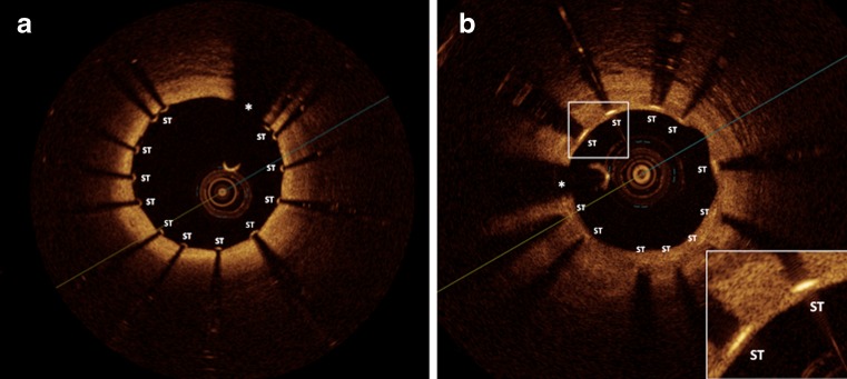
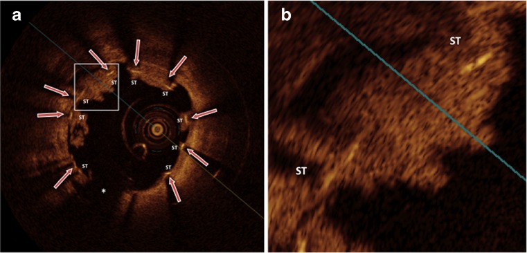
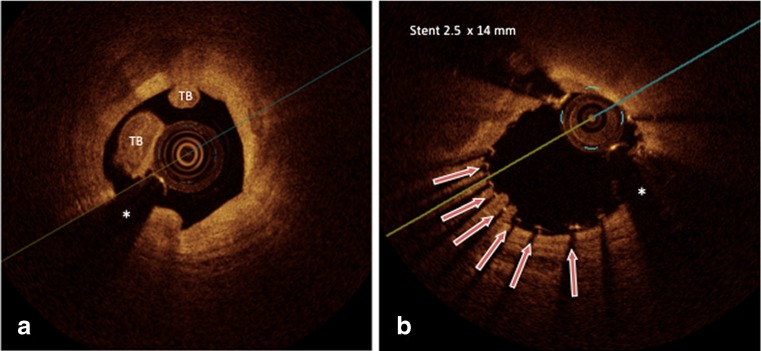
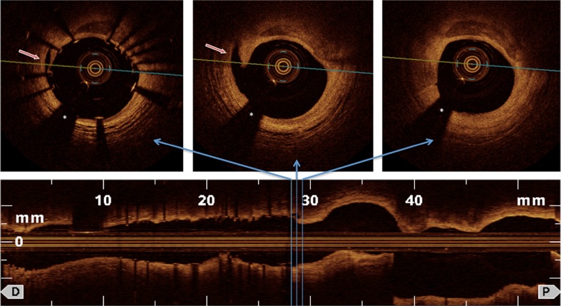
Optical coherence tomography (OCT) is a high-resolution imaging technique that is increasingly used for intracoronary imaging to characterize coronary atherosclerotic plaques and vascular responses after coronary stent implantation. Introduction of optical frequency-domain imaging (OFDI; second generation OCT) has simplified practical use of this novel imaging modality resulting in a more widespread availability in interventional cardiology. Here we highlight recent insights into the acute and chronic vascular response after coronary stent implantation by OCT imaging. OCT provides cross-sectional images with approximately 10-fold higher resolution as compared to intravascular-ultrasound (IVUS), allowing for precise evaluation of tissue coverage and malapposition of coronary stent struts. More than 30 studies using OCT to compare vascular responses to different stents have now been reported. Recent studies have examined the relation between OCT-image characteristics and tissue composition around stent struts. OCT is used for evaluation of novel stent concepts, such as bioengineered stents and bioabsorbable stents, where it provides more accurate information than IVUS. While intracoronary OCT imaging is further developed, including faster 3D-OCT-image-reconstruction, larger OCT studies/registries with standardized analysis will provide more insights into clinical implications of observations from OCT-imaging after coronary stent implantation.
Figures
Similar articles
-
Coronary optical frequency domain imaging (OFDI) for in vivo evaluation of stent healing: comparison with light and electron microscopy.Eur Heart J. 2010 Jul;31(14):1792-801. doi: 10.1093/eurheartj/ehq168. Epub 2010 Jun 5. Eur Heart J. 2010. PMID: 20525979 Free PMC article.
-
Comparison of Stent Expansion Guided by Optical Coherence Tomography Versus Intravascular Ultrasound: The ILUMIEN II Study (Observational Study of Optical Coherence Tomography [OCT] in Patients Undergoing Fractional Flow Reserve [FFR] and Percutaneous Coronary Intervention).JACC Cardiovasc Interv. 2015 Nov;8(13):1704-14. doi: 10.1016/j.jcin.2015.07.024. JACC Cardiovasc Interv. 2015. PMID: 26585621
-
The diagnostic value of intracoronary optical coherence tomography.Herz. 2011 Aug;36(5):417-29. doi: 10.1007/s00059-011-3487-7. Herz. 2011. PMID: 21744151 Review.
-
Evaluation in 3 months duration of neointimal coverage after zotarolimus-eluting stent implantation by optical coherence tomography: the ENDEAVOR OCT trial.JACC Cardiovasc Interv. 2009 Dec;2(12):1240-7. doi: 10.1016/j.jcin.2009.10.006. JACC Cardiovasc Interv. 2009. PMID: 20129551 Clinical Trial.
-
The role of optical coherence tomography in coronary intervention.Korean J Intern Med. 2012 Mar;27(1):1-12. doi: 10.3904/kjim.2012.27.1.1. Epub 2012 Feb 28. Korean J Intern Med. 2012. PMID: 22403493 Free PMC article. Review.
Cited by
-
Dynamic Assessment of the Endothelialization of Tissue-Engineered Blood Vessels Using an Optical Coherence Tomography Catheter-Based Fluorescence Imaging System.Tissue Eng Part C Methods. 2015 Jul;21(7):758-66. doi: 10.1089/ten.TEC.2014.0345. Epub 2015 Jan 30. Tissue Eng Part C Methods. 2015. PMID: 25539889 Free PMC article.
References
LinkOut - more resources
Full Text Sources